
Argon Laser Eye Treatment in Istanbul: Retinal Laser for Tears and Diabetic Eye Disease
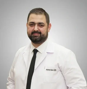
Argon laser eye treatment, also called retinal laser photocoagulation, uses a focused beam of light to seal retinal tears and to treat diabetic retinopathy and other retinal blood-vessel disease. In Istanbul it is planned and performed by Doç. Dr. Muhammet Derda Özer or Op. Dr. Aysan Bayram, after a full retina examination and scans. You know your surgeon and your written price before you travel, and a personal interpreter stays with you at every appointment.
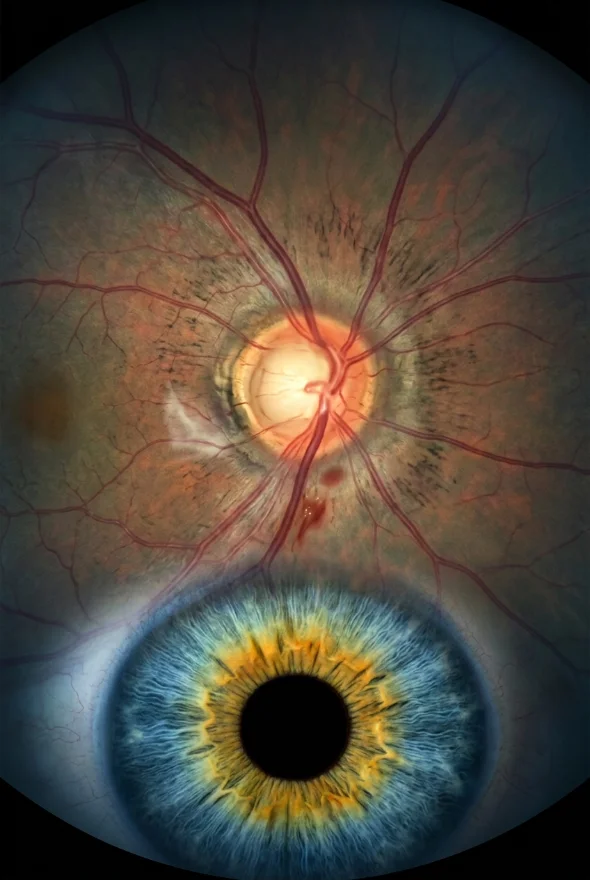
Argon laser eye treatment places tiny, controlled laser burns on the retina. Around a tear, they form a scar that welds the retina down (laser retinopexy). Across the outer retina, they calm the abnormal vessels of diabetic eye disease (panretinal photocoagulation). It takes about 20 minutes, under numbing drops, with no cut. In Istanbul, Doç. Dr. Özer or Op. Dr. Bayram treats you from {{£ price}} per eye; hotel and transfers are extra.
What is argon laser eye treatment?
Argon laser eye treatment is a retinal laser that turns light into heat inside the eye. Each laser spot is absorbed by the pigment layer under the retina and by blood, and the heat makes a small, deliberate scar. Doctors use these scars in two main ways:
- To glue the retina down. Spots placed in rows around a tear or weak area form scar tissue that acts as a barrier, so fluid cannot get under the retina and lift it off. This is laser retinopexy.
- To switch off the signal for abnormal vessels. In diabetic eye disease and some vein blockages, starved areas of retina release growth factors such as VEGF that make fragile new vessels grow. Spreading laser spots across the outer retina reduces that signal, and the new vessels shrink. This is panretinal photocoagulation (PRP), also called scatter laser.
Why it is called "argon". The first widely used retinal lasers ran on argon gas, which produces blue (488 nm) and green (514 nm) light. According to the American Academy of Ophthalmology's EyeWiki, argon green light is preferred near the centre of the retina, and today most clinics use newer 532 nm green lasers (frequency-doubled Nd:YAG) and pattern-scan lasers. Pattern lasers fire a grid of spots in 20 milliseconds each instead of the classic 100–200 milliseconds, which reduces heat spread and makes treatment more comfortable. Patients and many doctors still call all of these "argon laser", and the treatment principle is the same.
What is used for you: {{retinal laser platform name and wavelength}}, operated by your surgeon at a slit-lamp microscope or with a head-mounted (indirect) laser, in a fully equipped, Ministry of Health–licensed setting in Istanbul.
Before any laser, your surgeon examines the whole retina with your pupils dilated and usually takes an OCT scan of the macula, the central retina, to check for swelling. In diabetic eye disease, a dye test (fluorescein angiography) may be added to map leaking and starved areas.
Who needs retinal laser? Laser retinopexy indications and other uses
Retinal laser is used when the retina has a weak point that may tear or detach, or when diseased blood vessels threaten your sight. The main indications are:
| Condition | What the laser does | Typical treatment |
|---|---|---|
| Symptomatic horseshoe (flap) retinal tear | Seals the tear before fluid gets under it | Laser retinopexy, usually one session |
| Some asymptomatic tears, holes or lattice degeneration | Strengthens a weak area when your surgeon judges the risk worth treating | Laser retinopexy (often not needed; see below) |
| Proliferative diabetic retinopathy | Makes abnormal new vessels shrink | Panretinal photocoagulation (PRP), often in 1–4 sessions |
| Diabetic macular oedema outside the very centre | Seals leaking microaneurysms | Focal or grid laser, often alongside injections |
| Retinal vein occlusion with new vessels | Makes new vessels shrink | Scatter (PRP) or sectoral laser |
| Macular oedema after a branch vein occlusion | Reduces swelling | Grid laser in selected eyes |
Why a tear is treated. A retinal tear usually appears when the vitreous gel pulls away from the retina as we age (posterior vitreous detachment). An NHS patient leaflet reviewed in March 2026 states that this causes a tear in about 1 in 10 people who have a vitreous detachment. According to the American Academy of Ophthalmology (EyeWiki), 33–55% of untreated symptomatic horseshoe tears progress to retinal detachment. The same NHS leaflet says that laser treatment reduces this risk to less than 1 in 20.
Not every weak spot needs laser. The AAO's guidance is that asymptomatic atrophic or operculated holes rarely need treatment, and that acute horseshoe tears and tears after injury usually do. Your surgeon explains which group your eye is in.
Diabetic eye disease. In proliferative diabetic retinopathy, PRP has the longest track record of any treatment. In the US National Eye Institute's Diabetic Retinopathy Study, PRP cut the risk of severe vision loss by more than 50%: over 2 years, 6.4% of treated eyes lost vision versus 16.3% of untreated eyes (EyeWiki). For diabetic macular oedema, laser was the main treatment from the mid-1980s, when the ETDRS trial showed it roughly halved the risk of moderate vision loss (from 24% to 12% at 3 years). Since the 2000s, anti-VEGF injections have taken over for swelling that involves the centre, because they can improve vision, which laser rarely does (AAO EyeNet, 2021).
When it is an emergency, get treated at home first. If you suddenly notice new floaters, flashes of light or a dark curtain or shadow in your vision, the NHS advises getting help the same day. Do not wait for a trip abroad. Retinal laser in Istanbul makes sense for planned treatment: diabetic laser, laser your own eye doctor has recommended, a second opinion, or laser combined with another eye treatment while you are here.
Laser retinopexy vs laser photocoagulation: what is the difference?
Laser retinopexy is a form of laser photocoagulation. "Photocoagulation" describes the method (light that heats and seals tissue); "retinopexy" describes the purpose (fixing the retina in place). The table shows how the three main uses differ.
| Laser retinopexy | Panretinal photocoagulation (PRP) | Focal / grid laser | |
|---|---|---|---|
| Purpose | Seal a tear or weak area so the retina cannot detach | Make abnormal new vessels shrink | Reduce leakage and swelling at the macula |
| Where the spots go | A few rows around the tear only | Across the outer and mid retina, sparing the centre | Small spots near the centre |
| Number of spots | Small cluster | Classically about 1,000–2,000 per eye | Few |
| Sessions | Usually one | Often 1–4 | One or more, often with injections |
| Main trade-off | Small risk the tear does not fully seal | Some loss of side and night vision | Largely replaced by injections for centre-involving swelling |

Laser retinopexy settings: how the laser is applied
Your surgeon adjusts the settings to your eye in real time, aiming for a visible grey-white mark rather than a fixed number. As a guide, published settings are:
- Laser retinopexy for a tear: spot size 200–500 µm, duration 0.1–0.2 seconds, power raised until a grey-white burn appears. The tear is surrounded by at least three rows of spots, with no more than half a spot's width of untreated retina between them (EyeWiki).
- PRP for diabetic retinopathy: spot size 200–500 µm, about 100 milliseconds per spot on conventional lasers or 10–30 milliseconds on pattern lasers, aiming for grey rather than dense white burns (EyeWiki).
Laser can only reach the retina if the view is clear. If there is heavy bleeding in the eye or a dense cataract, freezing treatment (cryotherapy) or surgery may be used instead.
Argon laser eye treatment in Istanbul: price and what is included
With Laser Istanbul Clinic, retinal laser starts from {{£ price}} per eye. Laser retinopexy is usually a single session; panretinal laser for diabetic retinopathy may need several sessions, and your written quote states how many are included. Your surgeon confirms the exact price in writing after reviewing your eye report and scans, before you book anything.
Your treatment price includes:
- a full retina examination with dilating drops and the scans your surgeon needs (such as OCT)
- laser treatment by your named surgeon
- medication and eye drops
- follow-up checks in Istanbul with the same surgeon
- a personal interpreter, a dedicated coordinator and remote follow-up after you return home
Not included: flights, hotel and airport transfers. Hotel and transfers are a separate charge, and we arrange them for you if you ask.
If you live in the UK: urgent retinal tears and diabetic retinopathy are treated by NHS eye services, and you should use them for anything sudden. Patients who come to Istanbul usually do so for planned treatment, a second opinion or to combine retinal laser with other eye treatment in one trip.
Your retinal laser surgeons

Your retinal laser treatment, step by step
Free online review
Send your latest eye report, scans (OCT or retina photos) and diabetes details if relevant. A surgeon tells you whether laser is likely to be the right treatment and sends a written quote.
Book your dates
Plan {{number of days}} in Istanbul. Diabetic laser in several sessions may need a longer stay or a second visit; your surgeon plans this with you.
Examination
Your vision is measured and drops widen your pupils. Your surgeon examines the whole retina and takes the scans needed. If laser is not the right treatment, your surgeon tells you and explains the alternatives.
Laser treatment
Numbing drops are put in, and a special contact lens is placed on the eye to focus the laser. You sit at a slit-lamp microscope; for tears far out in the retina, you may lie back while the surgeon uses a head-mounted laser and presses gently on the eye. You see a series of bright flashes. It takes about 20 minutes.
Straight after
Your vision is dazzled and misty for a few hours. You go back to your hotel the same day; you must not drive that day.
Follow-up
For a tear, the surgeon checks that the laser has sealed it. NHS eye services typically review patients about two weeks after laser retinopexy; EyeWiki describes a check 1–2 weeks after treatment, again at 4–6 weeks, then at 3–6 months. Some of these checks can be done by your own eye doctor at home, with results shared with your surgeon. Your coordinator organises this.
Laser retinopexy recovery time
Most people go back to normal daily activities the next day, but the protective scar takes about two weeks to form fully. NHS patient leaflets state it takes about 14 days for the scar tissue to develop; EyeWiki reports maximal adhesion at roughly 7–10 days and a firm adhesion by 3 weeks.
-
First few hours
Dazzled, misty vision from the bright flashes; light sensitivity and glare outdoors. You may have a headache or an ache in the eye; paracetamol helps. No driving on the day of treatment.
-
Next day
Vision usually back to how it was before. Normal daily activities can usually resume.
-
First few days
Floaters can increase briefly, then settle. Some light sensitivity may continue.
-
Up to 5 days
Redness from the contact lens or from pressing on the eye settles on its own.
-
About 1–3 weeks
The laser scar matures and the retina is held firmly. Ask your surgeon which activities to pause until then.
-
1–2 weeks after laser
First check that the tear is sealed.
Laser does not remove floaters. Floaters you had before treatment are still there afterwards; with time they usually become less noticeable.
After PRP for diabetic retinopathy, you may feel dazzled for a few hours and should rest at home that day, according to NHS patient information; normal activities can usually resume the next day.
Laser retinopexy side effects and risks
Retinal laser is done without any incision, so, as NHS patient information notes, there is no risk of infection inside the eye. Side effects are usually mild and short-lived, but you should know all of them.
Common, short-lived side effects:
- Discomfort during treatment. Some people feel sharp, stabbing or aching pain, occasionally like a small electric shock. These sensations are unpleasant but do not mean harm inside the eye.
- Headache or a sore eye for a few hours.
- Dazzled, misty vision for a few hours, and glare or light sensitivity for a few days.
- Red eye from the contact lens, settling within about five days.
- More floaters for a few days.
Less common risks of laser retinopexy:
- The tear does not seal completely, and more laser is needed. Published failure rates for preventive treatment range from 0% to 22%, most often because the front edge of the tear was not fully covered (EyeWiki).
- Retinal detachment can still happen, although the risk is much lower than without treatment. Between 5% and 14% of people with a tear after a vitreous detachment develop further tears during long-term follow-up (AAO Preferred Practice Pattern, 2024).
- Bleeding inside the eye (vitreous haemorrhage), which normally clears by itself.
- An epiretinal membrane, a thin film on the retina that can blur or distort vision; usually mild, sometimes needing surgery.
- A laser spot in the wrong place, including the central vision, if the eye moves suddenly. This is why you are asked to keep very still.
Side effects specific to PRP for diabetic retinopathy:
- Reduced side (peripheral) vision and night vision, and sometimes colour vision. These changes are permanent.
- Driving. In the UK, the DVLA must be told if you have had laser treatment for diabetic retinopathy in both eyes; it may ask for a visual field test.
- Swelling of the macula and, rarely, temporary fluid around the choroid that blurs vision.
When to get help straight away: new or increasing floaters, flashing lights, a shadow or curtain across your vision, or a sudden drop in vision. These can signal a new tear or a detachment. Call your coordinator, and seek urgent eye care wherever you are.
Argon laser eye treatment: frequently asked questions
It is used to seal retinal tears and weak areas so the retina does not detach (laser retinopexy), and to treat diabetic retinopathy and retinal vein occlusion by shrinking abnormal blood vessels (panretinal photocoagulation). It is also used for some types of swelling at the macula, now often alongside injections. Your surgeon recommends it only after examining your retina.
Numbing drops are used, but they do not numb the inside of the eye completely. Many people feel brief sharp or aching sensations, especially during longer diabetic treatments, and some get a headache afterwards. Most find it manageable, and paracetamol helps afterwards.
The laser itself takes about 20 minutes. Allow extra time for the eye examination and for your pupils to widen beforehand. You go home the same day.
Vision is dazzled for a few hours and usually back to normal by the next day, when most people return to normal activities. The protective scar takes about 14 days to form fully. Your surgeon tells you which activities to avoid until then and checks the tear at about one to two weeks.
Common side effects are discomfort during treatment, a headache, dazzled vision for a few hours, light sensitivity, a red eye for up to five days and a brief increase in floaters. Less often, the tear does not fully seal and needs more laser, or bleeding or a thin film on the retina develops. A retinal detachment can still happen, but the risk is much lower than without treatment.
Laser retinopexy is one type of laser photocoagulation. Retinopexy places a few rows of spots around a tear to stop the retina detaching; panretinal photocoagulation spreads many spots across the outer retina to shrink abnormal vessels in diabetic eye disease. The laser is the same, but the purpose, number of spots and side effects differ.
No. Laser retinopexy is done to prevent a retinal detachment, not to clear floaters. Floaters you had before usually become less noticeable with time, and they can increase for a few days right after treatment.
Laser is applied through the pupil, without incisions or a gas bubble in the eye, so the flying ban that follows some retinal detachment operations does not apply. Your surgeon still checks your eye before you fly home and tells you when the follow-up check is due.
Not on the day of treatment, because your vision is dazzled and your pupils are dilated. In the UK, if you have had laser for diabetic retinopathy in both eyes, you must tell the DVLA, which may arrange a visual field test.
Both work. In the DRCR Retina Network's Protocol S trial, eyes treated with ranibizumab injections or with PRP had similarly good vision after five years; the injection group lost less side vision and developed less macular swelling, but needed regular injections. Laser is a one-off course, which can matter if you cannot attend frequent appointments. Your surgeon explains which suits your eyes.
Retinal laser is a routine, low-risk outpatient treatment when an experienced ophthalmologist examines the whole retina first and plans follow-up. Before you book, check your surgeon's name and qualifications, and how follow-up will work once you are home. If you have sudden new symptoms, get treated at home the same day rather than waiting to travel.
Other retina treatments in Istanbul
Retinal laser is one of several treatments for the retina. Which one you need depends on whether the retina is torn, detached, swollen or growing abnormal vessels.
| Retinal laser | Intravitreal (anti-VEGF) injections | Vitrectomy / retinal detachment surgery | |
|---|---|---|---|
| What it treats | Retinal tears, diabetic retinopathy, vein occlusion | Wet macular degeneration, diabetic macular oedema, vein occlusion | Retinal detachment, macular hole, bleeding that does not clear |
| How it is done | Light through the pupil; no incision | Medicine injected into the eye | Operation in theatre |
| Typical course | One session (tear) or 1–4 sessions (PRP) | Repeated injections, often monthly at first | Usually one operation |
| Recovery | Next day for most activities | Usually next day | Weeks; flying may be restricted if a gas bubble is used |
Intravitreal injections
Anti-VEGF injections reduce swelling and abnormal vessels, and can improve vision. They are often combined with laser.
Intravitreal injections (anti-VEGF eye injections)Retinal detachment surgery
If the retina has already lifted off, laser alone is usually not enough and surgery is needed.
Retinal detachment surgeryVitrectomy
Removes the vitreous gel to clear bleeding, release traction or repair the retina from inside the eye.
Vitrectomy in IstanbulMacular degeneration
Laser was once used for wet macular degeneration; today injections are the main treatment, and a magnifying macula lens can help some people with advanced disease.
Macular degeneration treatment and the Scharioth macula lensSources for the facts on this page
- EyeWiki (American Academy of Ophthalmology), Lasers (surgery): argon 488 nm blue / 514 nm green, absorption by RPE and haemoglobin, argon green preferred near the macula, 532 nm frequency-doubled Nd:YAG and 810 nm diode now dominate, pattern lasers 20 ms vs 100–200 ms, historical use for AMD before anti-VEGF. https://eyewiki.org/Lasers_(surgery)
- EyeWiki (AAO), Horseshoe or Flap Tear: 33–55% of symptomatic tears progress to detachment; laser settings 200–500 µm, 0.1–0.2 s, grey-white burns; at least three rows, ≤ half a spot size between burns; maximal adhesion ~7–10 days, firm adhesion at 3 weeks; prophylactic failure 0–22%; follow-up 1–2 weeks, 4–6 weeks, 3–6 months, then annually. https://eyewiki.org/Horseshoe_or_Flap_Tear
- EyeWiki (AAO), Panretinal Photocoagulation: ~1,000–2,000 burns (1,500–5,000 across 1–4 sessions in some protocols); 200–500 µm; 100 ms conventional, 10–30 ms short-duration; grey burns; DRS >50% reduction in severe visual loss (16.3% vs 6.4% at 2 years); Protocol S; permanent decreases in peripheral, colour and night vision; choroidal effusion. https://eyewiki.org/Panretinal_Photocoagulation
- American Academy of Ophthalmology, Posterior Vitreous Detachment, Retinal Breaks, and Lattice Degeneration PPP 2024: ~2% of acute PVDs without breaks develop breaks; 5–14% of patients with a break develop additional breaks; asymptomatic atrophic/operculated holes rarely need treatment, acute horseshoe and traumatic tears usually do. https://www.aao.org/education/preferred-practice-pattern/posterior-vitreous-detachment-retinal-breaks-latti
- Moorfields Eye Hospital NHS Foundation Trust, Laser retinopexy patient leaflet: scar ~14 days; slit-lamp contact-lens or indirect technique; stabbing/sharp/aching or electric-shock-like sensations; headache; redness settles within 5 days; misty vision for hours, normal by next day; no driving on the day; floaters not removed, may increase for a few days; no incision so no risk of intraocular infection; may not completely seal; detachment still possible; review ~2 weeks; warning symptoms. https://www.moorfields.nhs.uk/mediaLocal/zj1pq40t/laser-retinopexy.pdf
- University Hospitals of Leicester NHS Trust, Treating a tear or hole in your retina by laser (leaflet 1325, reviewed March 2026): tear in ~1 in 10 PVDs; about half of untreated tears detach, reduced to less than 1 in 20 with laser; ~20 minutes; ~14 days for scar; risks incl. vitreous haemorrhage, need for more laser, epiretinal membrane; no driving on the day; does not remove floaters. https://yourhealth.leicestershospitals.nhs.uk/library/musculoskeletal-specialist-surgery-mss/ophthalmology/2280-treating-a-tear-or-hole-in-your-retina-by-laser/file
- The Dudley Group NHS Foundation Trust, Argon laser pan-retinal photocoagulation (PRP) treatment: one to three sittings, about 20 minutes each, numbing drops and contact lens, up to 1,000 spots per session, reduced peripheral and night vision, tell DVLA, dazzled for a few hours, no driving that day, normal activities next day. https://www.dgft.nhs.uk/pil/argon-laser-pan-retinal-photocoagulation-prp-treatment/
- The Dudley Group NHS Foundation Trust, Argon laser retinopexy (photocoagulation) for retinal tear/break: redness 1–2 days, rare bleeding, treatment failure possible, review 2–4 weeks. https://www.dgft.nhs.uk/pil/argon-laser-retinopexy-photocoagulation-for-retinal-tear-break/
- NHS, Detached retina (retinal detachment): symptoms (floaters, flashes, dark curtain), same-day help via NHS 111, tears sealed with laser or freezing, risk factors. https://www.nhs.uk/conditions/detached-retina-retinal-detachment/
- NHS, Diabetic retinopathy: treatment: laser photocoagulation for proliferative diabetic retinopathy. https://www.nhs.uk/conditions/diabetic-retinopathy/treatment/
- GOV.UK, Eye conditions and driving: diabetic retinopathy with laser treatment must be reported to DVLA; conditions affecting both eyes; fine up to £1,000 for not reporting. https://www.gov.uk/diabetic-retinopathy-and-driving
- American Academy of Ophthalmology EyeNet, Elyasi N, Hemmati HD, Diabetic Macular Edema: Diagnosis and Management (May 2021): laser primary therapy from the mid-1980s after ETDRS; anti-VEGF since the 2000s can reverse vision loss, uncommon with laser; Protocol V. https://www.aao.org/eyenet/article/diabetic-macular-edema-diagnosis-and-management
- ETDRS result (focal/grid laser reduced moderate visual loss by ~50%, 24% → 12% at 3 years), as summarised in: Combination of Anti-VEGF and Laser Photocoagulation for Diabetic Macular Edema: A Review. J Ophthalmol 2017. https://pmc.ncbi.nlm.nih.gov/articles/PMC5350536/
- American Academy of Ophthalmology, Retinal Vein Occlusion: grid laser for macular oedema; scatter or sectoral PRP only once neovascularisation develops. https://www.aao.org/eye-health/diseases/retinal-vein-occlusion
- Gross JG, et al. (Writing Committee for the DRCR Retina Network). Five-Year Outcomes of Panretinal Photocoagulation vs Intravitreous Ranibizumab for Proliferative Diabetic Retinopathy. JAMA Ophthalmol 2018: similar visual acuity at 5 years; less visual field loss and less vision-impairing DME (22% vs 38%) with ranibizumab. https://pubmed.ncbi.nlm.nih.gov/30043039/
- Obeid A, et al. Outcomes of Eyes Lost to Follow-up with Proliferative Diabetic Retinopathy That Received Panretinal Photocoagulation versus Intravitreal Anti-VEGF. Ophthalmology 2019: more iris neovascularisation in the injection-only arm after loss to follow-up (background for the "one-off course" point; not quoted with numbers). https://www.aaojournal.org/article/S0161-6420(18)31079-0/fulltext
Find out if retinal laser is right for your eyes, free
Send us your latest eye report and any scans. Doç. Dr. Özer or Op. Dr. Bayram will tell you whether laser, injections or another treatment suits you, and what it would cost, with no obligation.
3 eye surgeons
Your case is reviewed by a named surgeon, not a salesperson. Reply within {{response time}}.
 Prof. Dr. Ahmet Satıcı
 Doç. Dr. Muhammet Derda Özer
 Op. Dr. Aysan Bayram