
- Home
- Knowledge centre
- Eye glossary A–Z
- Macular Degeneration (AMD): Types, Symptoms, Causes and Treatment
Macular Degeneration (AMD): Types, Symptoms, Causes and Treatment
Medically reviewed by {{surgeon}}, {{surgeon's title and specialty}}, on {{review date}}. Last updated {{date}}.
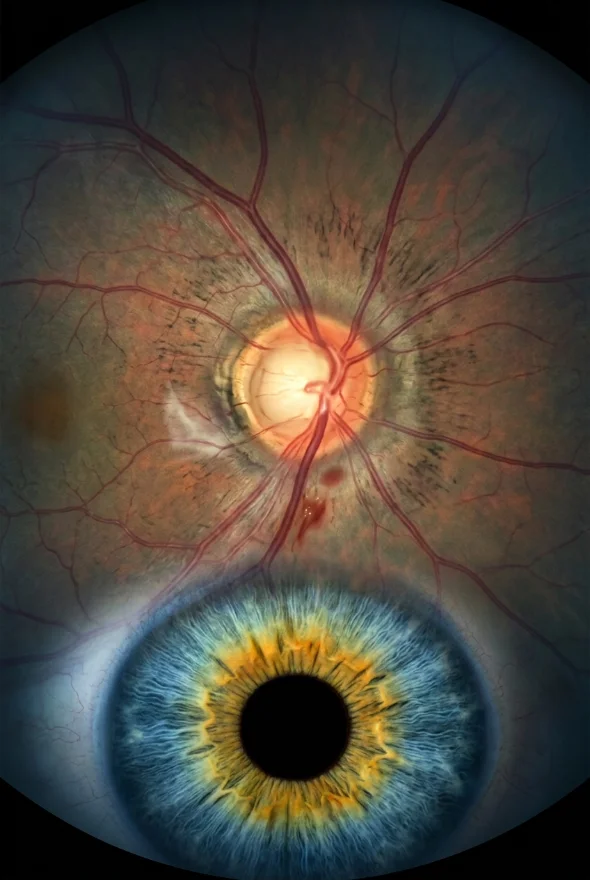
Macular degeneration (also age-related macular degeneration, AMD; noun): an eye disease in which ageing damages the macula, the small central part of the retina that gives sharp, straight-ahead vision. It blurs or distorts the centre of what you look at, which makes reading, driving and recognising faces hard, while side vision is kept. There are two forms, dry (the common one) and wet (less common, but faster and more serious), and wet macular degeneration needs urgent treatment with eye injections. In Istanbul, retina specialists Doç. Dr. Muhammet Derda Özer (Doç. = Associate Professor) and Op. Dr. Aysan Bayram assess macular disease.
Macular degeneration: key facts
- What it is
- Damage to the macula, the centre of the retina, caused by ageing; the National Eye Institute (NEI) calls it a leading cause of vision loss for older adults
- Who gets it
- Usually people over 50 (NHS); the NEI lists age 55 and over, white ethnicity, smoking and a family history as risk factors
- Main types
- Dry AMD (about 85–90% of cases) and wet AMD (about 10–15%, but the cause of most severe vision loss) (EyeWiki, American Academy of Ophthalmology)
- Typical symptoms
- Blurred or distorted central vision, straight lines that look wavy, a dark or empty patch in the middle, duller colours
- Does it cause blindness?
- It does not usually cause total blindness, because side vision is kept (NHS), but it can cause serious loss of central vision
- Diagnosis
- Dilated eye examination, optical coherence tomography (OCT) scan, Amsler grid, sometimes dye angiography
- Treatment
- Wet AMD: anti-VEGF injections into the eye. Intermediate dry AMD: AREDS2 vitamins. Late dry AMD: low-vision aids; two injections for geographic atrophy approved in the US
- Specialist
- Retina specialist (ophthalmologist); at Laser Istanbul Clinic, Doç. Dr. Muhammet Derda Özer and Op. Dr. Aysan Bayram

What is macular degeneration?
Macular degeneration is damage to the macula, the part of the retina that controls sharp central vision. The retina is the light-sensitive layer at the back of the eye. Its centre, the macula, is what you use to read, watch television, see fine detail and recognise a face. The rest of the retina gives you side (peripheral) vision.
When the macula is damaged, the middle of your vision blurs or goes missing, but the edges stay clear. The American Academy of Ophthalmology (AAO) gives a simple picture: you might see the numbers on a clock but not the hands. That is why the NHS says AMD does not cause total blindness but can make everyday tasks such as reading and recognising faces difficult.
AMD is common. The NEI estimates that 11 million people in the United States have it. A 2014 meta-analysis in The Lancet Global Health (Wong et al.) projected about 196 million people worldwide with AMD in 2020, rising to about 288 million by 2040.
What is the macula?
The macula is the small central area of the retina packed with the cells that detect fine detail and colour. It is what allows you to look straight at something and see it sharply. Damage to the macula therefore affects the part of vision you use most.
Types of macular degeneration: dry and wet
There are two forms of AMD, dry and wet, and they behave very differently.
| Dry AMD (atrophic) | Wet AMD (neovascular) | |
|---|---|---|
| What happens | Parts of the macula get thinner with age and tiny yellow deposits called drusen build up (AAO). In the late stage, patches of cells die (geographic atrophy) | New, abnormal blood vessels grow under the retina and leak fluid or blood, which can cause scarring (AAO) |
| How common | About 85–90% of AMD cases (EyeWiki) | About 10–15% of cases, but the cause of most severe vision loss (EyeWiki) |
| Speed | Usually slow, over years | Can worsen within weeks or months |
| Treatment | No treatment for early AMD; AREDS2 vitamins for intermediate AMD; injections for geographic atrophy in the US | Anti-VEGF injections into the eye, started quickly |
Dry AMD can turn into wet AMD, which is why people with dry AMD are asked to check their vision regularly and report changes.
The stages of dry AMD
Doctors stage AMD by the size of the drusen (EyeWiki, AAO):
- Early AMD: small or medium-sized drusen (under about 125 micrometres). There are usually no symptoms.
- Intermediate AMD: many medium drusen or any large drusen (125 micrometres or more). Symptoms may still be absent or mild.
- Late (advanced) AMD: geographic atrophy (dry form) or abnormal blood vessels, called choroidal neovascularisation (wet form). This is when central vision is lost.
In the Beaver Dam Eye Study, cited by EyeWiki, advanced AMD was found in about 1.6% of participants. In Wong et al.'s meta-analysis of people aged 45 to 85, early AMD affected about 8% and late AMD about 0.4%.
Macular degeneration symptoms
The main symptom of macular degeneration is a blurred, distorted or missing area in the centre of your vision. In the early stages there are often no signs at all (NEI), and changes can be so gradual that you do not notice them. According to the NHS and the NEI, symptoms can include:
| Symptom | What it looks like |
|---|---|
| Blurred or distorted central vision | Words on a page look fuzzy or a face looks blurred in the middle |
| Straight lines look wavy or crooked | Door frames, tiles and lines of text bend (doctors call this metamorphopsia) |
| A blurry or empty area near the centre | A dark or blank patch that can grow over time |
| Colours look less bright | Colours seem washed out |
| Difficulty reading, driving or recognising faces | Everyday tasks become harder |
| Hallucinations | Some people with significant sight loss see patterns, people or places that are not there; this is called Charles Bonnet syndrome and is linked to the damaged retina, not to mental illness |
AMD is painless and does not change the outward look of the eye (NHS). A red, painful eye points to a different problem.
Wet AMD can worsen quickly. If lines suddenly look wavy or a new dark patch appears, treat it as urgent. The NHS advises seeing an optician or calling NHS 111 urgently for sudden worsening of vision, a dark curtain or shadow moving across your vision, or a red and painful eye; the AAO says to call your ophthalmologist immediately if your Amsler grid changes. If this happens while you are abroad, see an eye doctor where you are; do not wait to travel.

Macular degeneration causes and risk factors
The exact cause of macular degeneration is not known, but ageing is the strongest risk factor. The NHS links AMD to ageing, smoking, high blood pressure, being overweight and a family history. In dry AMD the macula thins and drusen build up; in wet AMD new fragile blood vessels grow and leak.
The main risk factors listed by the AAO and the NEI are:
- Age: being over 50 (AAO) or 55 and older (NEI)
- Smoking: the main risk factor you can change. According to EyeWiki, current smokers are about twice as likely to lose vision from AMD as non-smokers, and people who stopped more than 20 years earlier showed no increased risk
- Family history of AMD
- White ethnicity
- High blood pressure, high cholesterol and heart disease
- Being overweight, and a diet high in saturated fat
You cannot change your age or your genes, but smoking, blood pressure, weight and diet are within your control.
How macular degeneration is diagnosed
AMD is diagnosed by examining the retina, and an OCT scan shows how severe it is. Eye doctors can check for AMD as part of a comprehensive dilated eye examination, in which drops widen the pupil so the retina and macula can be seen (NEI). The tests the AAO describes are:
- Amsler grid. A square grid of straight lines that reveals blurry or distorted spots. You look at it with one eye covered; wavy, blurry or missing lines are a warning sign.
- Dilated examination. Drops widen the pupil and the doctor looks at the retina with a special lens.
- Optical coherence tomography (OCT). A painless scan that creates a detailed cross-section picture of the retina. It shows drusen, thinning and fluid from wet AMD, and is also used to track how well treatment is working.
- Fluorescein angiography. Dye is injected into a vein in the arm, and photographs show abnormal blood vessels leaking in the macula.
- OCT angiography (OCTA). A newer scan that shows blood vessels without dye.
Checking your own eyes at home: the AAO describes using an Amsler grid daily. Hold it about 12 to 15 inches (30 to 38 cm) away, cover one eye, and look at the dot in the centre. Repeat with the other eye. Call your ophthalmologist immediately if lines look wavy, blurry or dim.
Macular degeneration treatment
Treatment depends on the type and stage of AMD, and no treatment brings back macular cells that have already died. The aim is to protect the vision you still have, or to help you use it better.
| Your diagnosis | Treatment | What it does |
|---|---|---|
| Early dry AMD | No specific treatment; healthy lifestyle and regular checks | Lowers the risk of progression |
| Intermediate dry AMD | AREDS2 vitamin and mineral supplement | In the original AREDS trial, the antioxidant-plus-zinc formula cut the risk of progression to advanced AMD by 25% (NEI) |
| Wet AMD | Anti-VEGF injections into the eye; in some cases laser or light treatment | Reduces leaking from abnormal vessels |
| Late dry AMD with geographic atrophy | Complement-inhibitor injections approved in the US | Slow the growth of the damaged area; they do not restore vision |
| Late AMD with lost central vision | Low-vision aids and rehabilitation | Make better use of remaining vision |
Wet AMD: anti-VEGF injections
VEGF is a growth signal that drives the abnormal blood vessels. Anti-VEGF medicines block it, and they are given through a very thin needle directly into the eye (AAO). They are the main treatment for wet AMD, and the NHS lists injections as one of the two approaches for wet AMD, alongside light treatment to destroy the vessels.
Wet AMD injections are not a one-off treatment: they are repeated over a long period. How often depends on the medicine and how your eye responds. EyeWiki notes that a newer medicine, faricimab, allowed most patients in trials to reach intervals of 3 months or more between injections. This is why many international patients start or continue injections near home and use a clinic abroad for a second opinion.
Dry AMD: AREDS2 vitamins
The NEI's AREDS2 formula contains vitamin C (500 mg), vitamin E (400 IU), zinc (80 mg), copper (2 mg), lutein (10 mg) and zeaxanthin (2 mg). It is for people with intermediate or advanced AMD; the NEI says the studies showed no benefit for people with early AMD or no AMD. The older AREDS formula contained beta-carotene, which increases the risk of lung cancer in current and former smokers, so smokers should avoid it. Ask your eye doctor before starting supplements.
Geographic atrophy: new injections
Until recently, advanced dry AMD had no treatment. The US FDA approved pegcetacoplan (Syfovre) on 17 February 2023 and avacincaptad pegol (Izervay) on 4 August 2023 for geographic atrophy (EyeWiki). Both slow the growth of the damaged area rather than restore vision. In trials, pegcetacoplan reduced lesion growth by about 36% at monthly dosing and avacincaptad pegol by about 35% (EyeWiki). Risks include a higher rate of conversion to wet AMD, infection, retinal detachment and raised eye pressure. In Europe, the European Medicines Agency refused marketing authorisation for pegcetacoplan in 2024, finding that slowing lesion growth did not lead to clinically meaningful benefits for patients and that the injections carried significant risks. Whether either medicine is available to you depends on where you live and on your doctor's advice.
Low-vision support
When central vision is lost, the AAO lists magnifying tools, handheld computers and other electronic aids, together with vision rehabilitation specialists to help you stay independent. In some selected patients with stable late AMD in both eyes, a surgical option is a small implanted magnifying lens for reading. It is a reading aid and does not treat the disease; it is described on our treatment page.
Treatment with us: your scans are reviewed by retina specialists Doç. Dr. Muhammet Derda Özer and Op. Dr. Aysan Bayram, who tell you which options suit your type and stage of AMD.
Can macular degeneration be prevented?
Macular degeneration cannot be fully prevented, but healthy habits can lower your risk and slow it down. The NEI and the AAO recommend:
- Not smoking. Stopping smoking is the most effective change you can make.
- Eating well. Dark leafy greens, yellow fruit and vegetables and fish are recommended; limit saturated fat.
- Staying active and keeping a healthy weight.
- Controlling blood pressure and cholesterol.
- Protecting your eyes from UV light.
- Regular eye examinations, especially after 50, so changes are found early.
If you already have intermediate AMD, ask your doctor whether AREDS2 supplements are right for you.

When to see a doctor about macular degeneration
Seek urgent same-day care (optician, eye emergency service or NHS 111 in the UK) if:
- straight lines suddenly look wavy or bent
- a new dark or blank patch appears in the centre of your vision
- your vision suddenly gets worse, or a dark curtain or shadow moves across it
- your eye is red and painful
Book a routine eye examination if:
- you are over 50 and have not had a dilated retinal examination
- reading is becoming harder, even with new glasses
- colours look duller or you need more light than before
- you have a family history of AMD, or you smoke
If you have already been diagnosed with AMD, check each eye daily with an Amsler grid and keep every appointment, because treatment of wet AMD works best when it starts early. If you want a second opinion on your treatment, you can send your OCT scans and eye report for review before you travel.
Frequently asked questions
Macular degeneration, or age-related macular degeneration (AMD), is an eye disease in which ageing damages the macula, the centre of the retina. It blurs the middle of your vision, making reading and recognising faces hard, but it leaves side vision intact.
Dry AMD is the common form, about 85–90% of cases: the macula thins and drusen build up, usually slowly. Wet AMD is less common, about 10–15%, but causes most severe vision loss: abnormal blood vessels leak under the retina and vision can worsen within weeks or months. Wet AMD is treated with anti-VEGF injections.
Early AMD often causes no symptoms. Later signs include blurred or distorted central vision, wavy straight lines, duller colours and a dark or empty area near the centre. A routine dilated eye examination can find the disease before you notice anything.
It does not usually cause total blindness, because side vision is kept, but it can cause serious loss of central vision that makes reading, driving and recognising faces difficult. Early treatment of wet AMD helps protect the sight you have.
No. There is no cure, and no treatment restores macular cells that have already died. Treatment aims to slow the disease or protect remaining vision: injections for wet AMD, AREDS2 vitamins for intermediate dry AMD, and low-vision aids for late AMD.
The exact cause is not known. Ageing is the strongest risk factor, and smoking, a family history, high blood pressure, being overweight and a diet high in saturated fat also raise the risk. Current smokers are about twice as likely to lose vision from AMD as non-smokers.
By a dilated eye examination and an OCT scan, which shows the layers of the macula. An Amsler grid, and sometimes dye or dye-free angiography, can add information about distortion and abnormal blood vessels.
Not entirely, but not smoking, eating leafy greens and fish, staying active, controlling blood pressure and cholesterol and protecting your eyes from UV light can lower your risk. AREDS2 vitamins can slow intermediate AMD.
For geographic atrophy, the US FDA approved pegcetacoplan (February 2023) and avacincaptad pegol (August 2023); they slow growth of the damaged area but do not restore vision. The European Medicines Agency refused pegcetacoplan in 2024, so availability depends on your country.
Sources for the facts on this page
- US National Eye Institute (NEI). Age-Related Macular Degeneration (AMD). https://www.nei.nih.gov/learn-about-eye-health/eye-conditions-and-diseases/age-related-macular-degeneration
- NHS. Age-related macular degeneration (AMD). https://www.nhs.uk/conditions/age-related-macular-degeneration-amd/
- NHS. AMD: symptoms. https://www.nhs.uk/conditions/age-related-macular-degeneration-amd/symptoms/
- NHS. AMD: treatment. https://www.nhs.uk/conditions/age-related-macular-degeneration-amd/treatment/
- American Academy of Ophthalmology (AAO). What Is Age-Related Macular Degeneration? (symptoms, causes, diagnosis, prevention). https://www.aao.org/eye-health/diseases/amd-symptoms
- American Academy of Ophthalmology. AMD treatment. https://www.aao.org/eye-health/diseases/amd-treatment
- EyeWiki (AAO). Age-Related Macular Degeneration. https://eyewiki.org/Age-Related_Macular_Degeneration
- EyeWiki (AAO). Geographic Atrophy. https://eyewiki.org/Geographic_Atrophy
- NEI. About the Age-Related Eye Disease Studies (AREDS and AREDS2). https://www.nei.nih.gov/research/clinical-trials/age-related-eye-disease-studies-aredsareds2/about-areds-and-areds2
- Wong WL, Su X, Li X, et al. Global prevalence of age-related macular degeneration and disease burden projection for 2020 and 2040: a systematic review and meta-analysis. The Lancet Global Health 2014. PubMed: https://pubmed.ncbi.nlm.nih.gov/25104651/
- European Medicines Agency. Syfovre (pegcetacoplan), refusal of marketing authorisation. https://www.ema.europa.eu/en/medicines/human/EPAR/syfovre
Diagnosed with macular degeneration, or noticing distortion in your vision?
Send us your OCT scans and eye report. Our retina specialists, Doç. Dr. Muhammet Derda Özer and Op. Dr. Aysan Bayram, will review them and tell you which options may suit your eyes, with no obligation.
3 eye surgeons
Your case is reviewed by a named surgeon, not a salesperson. Reply within {{response time}}.
 Prof. Dr. Ahmet Satıcı
 Doç. Dr. Muhammet Derda Özer
 Op. Dr. Aysan Bayram