
- Home
- Knowledge centre
- Eye glossary A–Z
- Eye Stroke (Retinal Artery Occlusion): Symptoms, Causes and Treatment
Eye Stroke (Retinal Artery Occlusion): Symptoms, Causes and Treatment
Medically reviewed by {{surgeon}}, {{surgeon's title and specialty}}, on {{review date}}. Last updated {{date}}.
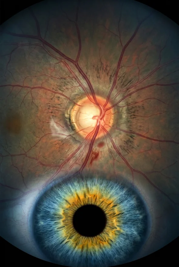
for eye stroke treatment on the NHS, the route is emergency care, not a routine appointment. An eye stroke, medically a retinal artery occlusion, is a blockage of the artery feeding the retina. It causes sudden, painless loss of vision in one eye and is treated as a form of stroke. Our retina specialists, Doç. Dr. Muhammet Derda Özer and Op. Dr. Aysan Bayram, see patients once the emergency has been dealt with.
Eye stroke (informal term, also "retinal stroke"): a sudden blockage of an artery supplying the retina, called a retinal artery occlusion (RAO). Without blood, the retina is starved of oxygen and the nerve cells that carry sight to the brain are damaged within hours. It is called an eye stroke because it works like a stroke in the brain: a clot or fragment of plaque lodges in a small artery. The American Heart Association describes central retinal artery occlusion as a form of acute ischaemic stroke and a warning of further stroke and heart problems.
Doç. is the Turkish title for Associate Professor.
Eye stroke (retinal artery occlusion): key facts
- Medical name
- Retinal artery occlusion: central (CRAO) or branch (BRAO)
- Main symptom
- Sudden, painless loss of vision in one eye, or of part of the visual field
- Who gets it
- Mostly older adults, more often men; also people with high blood pressure, diabetes, high cholesterol, heart rhythm problems or who smoke
- How common
- About 1.9 new cases of central occlusion per 100,000 people a year in the United States, rising to about 10 per 100,000 in people over 80
- Is it an emergency?
- Yes. It needs immediate assessment, ideally at a hospital with stroke services
- Can vision come back?
- Often only partly, and in many people very little. Treatment options are limited and still being tested
- Linked risk
- Higher risk of stroke and heart attack afterwards, so a full vascular check is needed
- Specialist
- Emergency, stroke and eye teams at the time; later, a retina specialist (ophthalmologist). At Laser Istanbul Clinic: Doç. Dr. Muhammet Derda Özer or Op. Dr. Aysan Bayram
What is an eye stroke? Central, branch and "mini" eye strokes
An eye stroke is not one condition but a group of related blockages. The retina gets its blood from the central retinal artery, which divides into branches. Where the blockage sits decides how much vision you lose.
| Type | What is blocked | What it usually feels like |
|---|---|---|
| Central retinal artery occlusion (CRAO) | The main artery to the retina | Severe, sudden loss of vision across the whole eye |
| Central occlusion with cilioretinal artery sparing | The main artery, but a small extra vessel (present in about one in three eyes) still feeds the macula | Central vision can be much better preserved; the surrounding field is still lost |
| Branch retinal artery occlusion (BRAO) | One branch of the artery. According to EyeWiki, about 38% of all retinal artery occlusions | A dark patch or missing area in part of the field of vision, usually less severe than CRAO |
| Transient occlusion ("mini eye stroke") | A blockage that clears by itself | Vision goes, then returns. Doctors call this transient monocular vision loss or amaurosis fugax |
Is a mini eye stroke serious? Yes. A transient episode means the same artery disease or clot source is present, and it is a warning that a full stroke or a permanent eye stroke can follow. The NHS describes a mini stroke (TIA) in the brain as a warning sign of a possible full stroke and says you need hospital assessment even if the symptoms disappear. The same principle applies when vision goes in one eye and then comes back.

Eye stroke symptoms
The symptoms of an eye stroke are sudden, painless loss of vision in one eye. The loss typically develops over seconds. Look for:
- sudden loss of all vision in one eye, or a dark shadow or "curtain" across part of it
- vision that is blurred, dim or greyed-out in one eye, with no pain
- a blind patch in one eye, as in a branch occlusion
- colours looking faded in that eye
- a brief episode of vision loss in one eye that then clears (mini eye stroke symptoms are the same, but vision returns)
- other stroke signs at the same time: face drooping, arm weakness or slurred speech
Eye stroke is painless, which is why it can be mistaken for something minor. A painful red eye points to other problems, such as acute glaucoma or inflammation, which also need urgent care.
Eye stroke pictures: what a doctor sees
An eye doctor looking at the back of the eye often sees a pale, cloudy-white retina with a small red "cherry-red spot" at the macula, the centre of the retina. A review of the evidence reports this in up to about 90% of central occlusions. The retinal arteries may look thin, and the blood in them may break into segments, sometimes called "box-carring". On OCT (optical coherence tomography) scans, the inner retina looks swollen and bright at first, and thins in the weeks afterwards.
Eye stroke causes and risk factors
Most eye strokes are caused by an embolus, a clot or fragment of plaque that travels from the heart or the carotid artery in the neck and lodges in the retinal artery. Emboli tend to stick where the artery narrows as it enters the optic nerve.
| Cause | How it blocks the artery |
|---|---|
| Carotid artery disease | Plaque in the neck artery sheds fragments that travel to the eye (a "Hollenhorst plaque" is a cholesterol fragment seen in the retinal artery) |
| Heart conditions | Atrial fibrillation (an irregular heartbeat), heart valve disease or a hole in the heart can send clots to the eye |
| Giant cell arteritis (temporal arteritis) | Inflammation of the arteries, mostly over the age of 50; about 5% of central occlusions are of this inflammatory (arteritic) type |
| High blood pressure, diabetes, high cholesterol, smoking | Damage the artery walls and raise the risk of clots |
| Blood-clotting disorders | Looked for particularly in patients under 50 without the usual risk factors |
| Other causes | Some medical procedures and dialysis, injury, certain infections and, rarely, spasm of the artery |
Giant cell arteritis matters because it is treatable and the other eye is at risk. The NHS lists severe headaches, tenderness at the temples, jaw pain when eating or talking, and loss of vision among its signs, and warns that it "can lead to serious problems like stroke and blindness if not treated quickly". If you are over 50 and have these symptoms with sudden vision loss, say so at once: high-dose steroids are started immediately to protect the other eye, while tests continue.
How serious is an eye stroke? Can you die from it?
An eye stroke is serious for two reasons: the eye itself, and what it says about the rest of your blood vessels.
For the eye: blood flow to the retina must be restored quickly. Clinical observations and animal studies suggest irreversible damage can occur after roughly 100 to 240 minutes of blockage, and some animal studies show damage to retinal cells within 12 to 15 minutes. In a classic central occlusion, vision in the first week stays the same in about two in three cases, improves in about one in five and worsens in about one in eight. Only a minority of patients regain useful central vision.
For the body: a central occlusion is a stroke of the retina and a warning sign for the brain and heart.
- A meta-analysis cited in a 2024 review found that about 30% of people with CRAO had a new ischaemic stroke on brain MRI within 7 days of symptoms, often without brain symptoms.
- In a 2026 UK study of 566 patients at a London eye hospital, people with CRAO had a nearly threefold higher risk of later stroke or heart attack than matched controls. This was not seen after branch occlusion or transient vision loss, though the numbers were small. Atrial fibrillation was newly found in 6% of CRAO patients within a year.
- A 2026 analysis of about 92,000 patients with retinal vascular occlusion compared them with patients who had a TIA. Central retinal artery occlusion carried a higher risk of major cardiovascular events (hazard ratio 1.13) and death (1.23) than TIA.
Can you die from an eye stroke? The eye stroke itself does not usually kill, but the vascular disease behind it can. The 2026 analysis above found higher all-cause mortality after CRAO than after TIA. This is why you need stroke-style tests, not just an eye check.
Can an eye stroke lead to a brain stroke? Yes. The risk is highest in the first days and weeks, which is why urgent investigation and preventive treatment matter even if your vision recovers.
How is an eye stroke diagnosed?
The diagnosis is usually made by examining the eye, then looking for the source of the clot.
Eye examination
Visual acuity, pupil reaction to light, and a dilated look at the retina.
OCT retinal scan
A quick, painless scan that shows swelling and damage in the retina.
Fluorescein angiography
A dye test, used in some cases, that shows delayed filling of the retinal artery.
Blood tests
Blood sugar, cholesterol, clotting tests, and ESR and CRP inflammation markers if giant cell arteritis is possible.
Brain and neck imaging
CT or MRI of the head, and imaging of the carotid arteries (ultrasound or angiography) to look for narrowing.
Heart tests
An ECG and heart rhythm monitoring for atrial fibrillation, and an ultrasound scan of the heart (echocardiogram).
Eye stroke treatment: what can be done?
There is currently no treatment proven to restore vision after an eye stroke. EyeWiki states that no evidence-based treatment has yet been shown to give a visual benefit. A 2023 Cochrane review of six small randomised trials (223 people in total) found no clear advantage of any treatment over observation or standard measures, though the evidence was of low certainty.
| Approach | What it is | Evidence |
|---|---|---|
| Clot-busting drug (thrombolysis) | Alteplase or a similar drug through a drip, as in brain stroke | The French THEIA trial (Lancet Neurology, 2025) compared alteplase with aspirin within 4.5 hours in 70 patients. Among those with results, 66% on alteplase and 48% on aspirin improved by a set amount at one month, but the difference was not statistically significant and the trial was probably too small. One symptom-free brain bleed was reported in the alteplase group. More trials are under way |
| Eyeball massage, lowering eye pressure, draining fluid from the front of the eye | Tries to push the clot onward | Used for decades; reviews describe minimal proven benefit, and massage carries a risk of moving the clot further along the vessel |
| Special oxygen mixtures or hyperbaric oxygen | Tries to supply oxygen to starved tissue | Limited, low-quality evidence |
| Intra-arterial thrombolysis | Drug delivered by catheter to the eye's artery | EyeWiki reports it was abandoned because of brain bleeding without visual benefit |
| Steroids for giant cell arteritis | High-dose steroids when inflammation is suspected | Essential if present, to protect the other eye |
| Secondary prevention | Treating blood pressure, diabetes and cholesterol; antiplatelet medicines; stopping smoking; treating carotid disease or atrial fibrillation | The most important long-term treatment: its aim is to prevent a brain stroke or heart attack |
Is an eye stroke curable? The blocked artery may reopen over days to weeks, but the retinal damage done by then is usually permanent. Low-vision support can help people adapt, and later complications inside the eye can be treated.
Eye stroke treatment NHS patients can expect: what happens in hospital
We could not find a dedicated NHS patient page on eye stroke, so this section describes the guidance that does exist. The NHS stroke pages list "blurred vision or loss of sight in 1 or both eyes" among stroke warning signs and tell you to call 999 and not to drive yourself. The American Heart Association says acute CRAO is a medical emergency and notes that management varies considerably between hospitals and specialities. In practice, expect an assessment by emergency doctors and often a stroke or eye team, the tests above, and a plan to prevent further strokes. Whether thrombolysis is offered depends on the hospital and on how quickly you arrive, so ask the team directly. Outside the UK the same approach applies: go straight to an emergency department with stroke services, not to a routine clinic.

How long does an eye stroke last?
A mini eye stroke clears by itself, but a true eye stroke usually leaves lasting damage. A transient episode, by definition, resolves; for a TIA in the brain the NHS says symptoms typically last minutes to hours. In a true retinal artery occlusion the artery may reopen over a week or more, but vision lost in the first hours often does not fully return. Some people see slight improvement over the first days, but the figures above show most stay the same. Do not wait to find out which kind you have: at the start they feel identical.
Outlook and recovery
- Central occlusion with a spared cilioretinal artery: EyeWiki reports that vision usually recovers to 20/50 or better in more than 80% of eyes.
- Branch occlusion: EyeWiki reports 20/40 or better in about 80% of eyes; one series of permanent branch occlusions put it at 89%.
- Classic central occlusion: severe, usually permanent loss of vision, with a reduced field of vision.
- Complications: abnormal new blood vessels can later grow in the eye; EyeWiki lists laser treatment and anti-VEGF injections as options. Your eye doctor will check for this at follow-up.
- Day-to-day life: losing sight in one eye affects depth perception, driving and reading. Ask about vision aids and driving rules in your country.

When to get help
Call an ambulance now (999 or your local number) if:
- you lose vision in one eye suddenly, even if it comes back
- vision loss comes with face drooping, arm weakness, slurred speech, confusion or severe headache
- you are over 50 with new headache, scalp or temple tenderness or jaw pain when chewing, plus any change in vision
Same-day eye or emergency care: a new dark patch or shadow in one eye, or a painful red eye with blurred vision.
Routine follow-up (with your GP or eye doctor): after an eye stroke, ongoing checks of blood pressure, cholesterol, diabetes and heart rhythm, and regular retinal checks.
What our retina doctors can do
An eye stroke cannot be treated by travelling abroad: it must be assessed in the first hours, where you are. What we offer is the stage after that: a specialist retinal assessment including OCT scanning, a second opinion on your scans and reports, follow-up for complications in the eye, and advice on other retinal conditions. Retina care is led by Doç. Dr. Muhammet Derda Özer, whose specialities include retina and uveitis, together with Op. Dr. Aysan Bayram, who treats retinal disease and speaks English. Retinal surgery such as vitrectomy and retinal detachment repair treats different conditions and is not a treatment for an acute eye stroke.
Frequently asked questions
An eye stroke is a sudden blockage of an artery supplying the retina, called a retinal artery occlusion. It causes sudden, painless loss of vision in one eye and is treated as an emergency, like a stroke in the brain.
Sudden, painless loss of vision in one eye, or a dark shadow or blind patch in part of it. Colours may look faded. Sometimes vision goes and then returns, which is a mini eye stroke and just as urgent.
We could not find a dedicated NHS patient page for eye stroke. NHS stroke guidance says to call 999 for loss of sight in one or both eyes. Treatment then depends on the hospital: assessment by emergency, stroke and eye teams, tests to find the cause, and preventive medicines. Clot-busting drugs are still being tested.
No treatment has been proven to restore lost vision. Vision recovers well only in some patients, especially when a small spared vessel supplies the centre of the retina. Treating the cause is crucial to prevent a second event.
The eye stroke itself is not usually fatal, but it signals a higher risk of stroke, heart attack and death from vascular disease. A 2026 analysis found a higher risk of death after central retinal artery occlusion than after TIA.
Yes. About 30% of people with central retinal artery occlusion were found to have new ischaemic changes on a brain MRI within a week, and the risk of a later stroke or heart attack is higher. Prompt investigation and treatment of the cause reduce that risk.
A mini eye stroke clears on its own. A true occlusion causes lasting damage; some improvement is possible in the first days, but most people do not regain full vision.
Mini eye stroke symptoms are the same as a full eye stroke, but vision returns on its own. It is a warning of stroke or permanent vision loss and needs emergency assessment the same day.
Sources for the facts on this page
- NHS. Stroke: symptoms. https://www.nhs.uk/conditions/stroke/symptoms/
- NHS. Transient ischaemic attack (TIA). https://www.nhs.uk/conditions/transient-ischaemic-attack-tia/
- NHS. Temporal arteritis (giant cell arteritis). https://www.nhs.uk/conditions/giant-cell-arteritis/
- EyeWiki (AAO). Central Retinal Artery Occlusion. https://eyewiki.org/Central_Retinal_Artery_Occlusion
- EyeWiki (AAO). Branch Retinal Artery Occlusion. https://eyewiki.org/Branch_Retinal_Artery_Occlusion
- Mac Grory B, Schrag M, Biousse V, et al. Management of Central Retinal Artery Occlusion: A Scientific Statement From the American Heart Association. Stroke 2021. PubMed: https://pubmed.ncbi.nlm.nih.gov/33677974/
- Dagra A, Lucke-Wold B, et al. Central Retinal Artery Occlusion: A Review of Pathophysiological Features and Management. Stroke: Vascular and Interventional Neurology 2024. https://pmc.ncbi.nlm.nih.gov/articles/PMC12778453/
- Préterre C, et al. Intravenous alteplase versus oral aspirin for acute central retinal artery occlusion within 4.5 h of severe vision loss (THEIA). Lancet Neurology 2025. https://doi.org/10.1016/s1474-4422(25)00308-4
- Lin JC, Song S, Ng SM, Scott IU, Greenberg PB. Interventions for acute non-arteritic central retinal artery occlusion. Cochrane Database of Systematic Reviews 2023. https://pmc.ncbi.nlm.nih.gov/articles/PMC9885744/
- Huemer J, et al. Risk of cardiovascular events and death following retinal arterial occlusion and transient monocular visual loss. Scientific Reports 2026. https://doi.org/10.1038/s41598-026-58854-8
- Chuang CW, Hsieh CY, Sung SF. Risk of Major Adverse Cardiovascular Events After Retinal Vascular Occlusions Versus Transient Ischemic Attack. Journal of the American Heart Association 2026. https://doi.org/10.1161/jaha.126.049229
- Roskal-Wałek J, et al. Therapeutic Strategies for Retinal Artery Occlusion: A Literature Review. Journal of Clinical Medicine 2024. https://pmc.ncbi.nlm.nih.gov/articles/PMC11595154/
Had an eye stroke or sudden vision loss? Want a retina specialist's second opinion afterwards?
Once you are safe and have been seen urgently, send us your scans and reports. A retina specialist will review them and tell you what the findings mean for your eye, with no obligation.
3 eye surgeons
Your case is reviewed by a named surgeon, not a salesperson. Reply within {{response time}}.
 Prof. Dr. Ahmet Satıcı
 Doç. Dr. Muhammet Derda Özer
 Op. Dr. Aysan Bayram