
- Home
- Knowledge centre
- Eye glossary A–Z
- Diabetic Retinopathy: What It Is, Stages, Symptoms and Treatment
Diabetic Retinopathy: What It Is, Stages, Symptoms and Treatment
Medically reviewed by {{surgeon}}, {{surgeon's title and specialty}}, on {{review date}}. Last updated {{date}}.
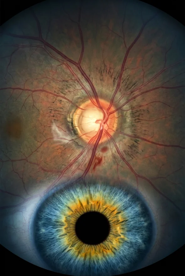
Diabetic retinopathy (noun): diabetic retinopathy is damage to the tiny blood vessels of the retina, the light-sensing layer at the back of the eye, caused by long-term high blood sugar in people with diabetes. The vessels can leak, close off or be replaced by fragile new ones that bleed, which can blur vision and, if untreated, lead to permanent sight loss. Early on it usually causes no symptoms, so regular eye screening matters. At Laser Istanbul Clinic, retina care is led by Doç. Dr. Muhammet Derda Özer and Op. Dr. Aysan Bayram.
Diabetic retinopathy: key facts
- What it is
- A complication of diabetes that damages the blood vessels of the retina
- Who gets it
- People with type 1, type 2 or gestational diabetes; the risk rises the longer diabetes lasts
- How common
- The US National Eye Institute (NEI) says that over time more than half of people with diabetes will develop it
- Main stages
- Non-proliferative (early, with leaky vessels) and proliferative (advanced, with new abnormal vessels)
- Main threat to sight
- Swelling of the macula (diabetic macular oedema) and bleeding or scarring in advanced disease
- Early symptoms
- Usually none
- Detection
- Dilated eye examination or retinal photography, often with an OCT scan
- Treatment
- Blood sugar and blood pressure control, anti-VEGF injections, laser, steroid treatment, and vitrectomy surgery for advanced disease
- Specialist
- Retina specialist (ophthalmologist); at Laser Istanbul Clinic, Doç. Dr. Muhammet Derda Özer or Op. Dr. Aysan Bayram

What causes diabetic retinopathy?
The cause is high blood glucose damaging the retina's small blood vessels over time. The American Academy of Ophthalmology (AAO) explains that these vessels can swell, leak, close off, or be replaced by abnormal new vessels, any of which can cause vision loss.
The retina needs a steady blood supply. When vessels leak, fluid collects in the retina. When they close, parts of the retina are starved of oxygen, and the retina responds by growing new vessels. These new vessels are weak, bleed easily and form scar tissue, which is why advanced disease is so damaging.
Who is at risk of diabetic retinopathy?
Anyone with diabetes can develop it. The main risk factors named by the NHS and NEI are:
- Long-standing diabetes. The risk grows with every year of diabetes. According to EyeWiki (AAO), after 20 years of diabetes, about 99% of people with type 1 and about 60% of people with type 2 have some retinopathy.
- High blood sugar that is poorly controlled.
- High blood pressure.
- High cholesterol.
- Pregnancy. The NEI notes that women with diabetes who become pregnant are at high risk of developing retinopathy, so they need closer eye checks.
- Smoking. The NHS lists avoiding smoking as part of protecting your sight.
Diabetes is common and rising: the World Health Organization (WHO) reports that the number of people living with diabetes rose from 200 million in 1990 to 830 million in 2022. The US Centers for Disease Control and Prevention (CDC) calls diabetic retinopathy the leading cause of blindness in working-age adults.
What are the stages of diabetic retinopathy?
Diabetic retinopathy has two main stages, and doctors grade the early stage further.
| Stage | What happens in the retina | Typical symptoms | Usual approach |
|---|---|---|---|
| Non-proliferative diabetic retinopathy (NPDR): mild to moderate | Small vessels leak; tiny bulges (microaneurysms), small haemorrhages and fluid appear | Often none | Monitoring, tighter control of blood sugar and blood pressure; the NHS says background or pre-proliferative retinopathy does not need treatment, only regular monitoring |
| Severe NPDR | Many more vessels are blocked, so large areas of retina are starved of oxygen. Doctors use the "4-2-1 rule" (EyeWiki): bleeding in all 4 quadrants of the retina, venous beading in 2 or more, or abnormal vessel segments (IRMA) in 1 or more | Often none | Close monitoring by a retina specialist; treatment may be considered |
| Proliferative diabetic retinopathy (PDR) | The retina grows new, fragile blood vessels that bleed into the vitreous gel or form scar tissue | Floaters, dark patches, sudden blurring; sometimes sudden vision loss | Laser, anti-VEGF injections, and surgery if there is heavy bleeding or scarring |
| Diabetic macular oedema (DME) | Fluid builds up in the macula, the central retina used for reading and faces. It can occur at any stage | Blurred or distorted central vision, trouble reading | Anti-VEGF injections, steroid treatment or laser |
The AAO says macular swelling is the most common reason people with diabetes lose vision. According to the NEI, about 1 in 15 people with diabetes will develop diabetic macular oedema over time.
In proliferative disease, scar tissue can pull on the retina and cause a tractional retinal detachment, and bleeding into the vitreous can block vision altogether.
What are the symptoms of diabetic retinopathy?
In the early stages, diabetic retinopathy usually causes no symptoms. The NHS, NEI and CDC all say the same: you can have it and see normally. This is why screening, not symptoms, is how most people are diagnosed.
As the condition advances, the AAO, NHS and CDC list these symptoms:
| Symptom | What it can mean |
|---|---|
| Blurred vision, or vision that goes in and out of focus | Macular swelling (DME) or bleeding |
| New floaters: dots, lines or dark shapes drifting in your vision | Bleeding into the vitreous gel (PDR) |
| Blank or dark areas in your vision | Bleeding, or damaged areas of retina |
| Poor night vision or difficulty in dim light | Widespread retinal damage |
| Faded or washed-out colours | Macular involvement |
| Sudden or severe vision loss | Heavy bleeding or retinal detachment: urgent |
Symptoms often affect both eyes, but one eye can be worse than the other.
How is diabetic retinopathy diagnosed?
A retina specialist or screening service looks at the back of the eye after widening the pupils with drops. The usual tests are:
Dilated eye examination or retinal photography
Drops widen your pupil so the doctor, or a special camera, can see the retina. In the NHS England diabetic eye screening programme, an appointment takes about 30 minutes and includes a letter chart, dilating drops and photographs. Your sight may be blurry for a few hours afterwards, so do not drive.
Optical coherence tomography (OCT)
A scan, similar to an ultrasound that uses light, shows the thickness of the retina in cross-section. It is the key test for detecting and measuring macular swelling.
Fluorescein angiography or OCT angiography
A yellow dye (fluorescein) injected into an arm vein shows leaking, blocked or abnormal vessels as it passes through the retina. OCT angiography can show vessels without dye, but with less detail. The NEI says your doctor may use angiography if severe retinopathy or DME is suspected.
Before any treatment at Laser Istanbul Clinic, your surgeon examines the retina and reviews your OCT and retinal photographs. You can send your existing scans for review before you travel.
How is diabetic retinopathy treated?
Treatment depends on the stage and on whether the macula is affected. Mild disease needs monitoring and good diabetes control; advanced disease needs treatment inside the eye. The NHS notes that it is not possible to cure sight loss already caused by diabetic retinopathy, which is why catching and treating it early matters.
| Treatment | What it does | Typically used for |
|---|---|---|
| Blood sugar, blood pressure and cholesterol control | Slows damage to the retinal vessels; the AAO notes that good sugar control can even bring some vision back | Every stage |
| Anti-VEGF injections (for example Avastin, Eylea, Lucentis) | Medicine injected into the eye blocks the growth factor that makes vessels leak and grow; the NEI says it can slow down or reverse diabetic retinopathy | Diabetic macular oedema and proliferative disease |
| Laser treatment (focal laser, or panretinal photocoagulation, PRP) | Seals leaking vessels, or treats the outer retina so abnormal new vessels shrink; often several sessions | Proliferative disease; some macular oedema |
| Steroid treatment (injection or slow-release implant) | Reduces swelling in the macula | Macular oedema that does not respond well to other treatment |
| Vitrectomy | Keyhole surgery removes blood and the vitreous gel, and removes scar tissue that pulls on the retina | Heavy vitreous bleeding, tractional retinal detachment |
Anti-VEGF injections or laser?
For macular oedema, anti-VEGF injections are the first-line treatment (EyeWiki). In the DRCR Protocol S trial, the anti-VEGF drug ranibizumab was found non-inferior to PRP for proliferative disease, so injections or laser may both be options. The AAO's 2024 Preferred Practice Pattern says that PRP remains an important treatment for PDR, and that when the macula is involved but vision is still good, treatment may reasonably be deferred until vision falls to 20/30 or worse. The choice depends on your eye, how reliably you can attend repeat injections, and your own doctor's advice. Injections are given after the eye is numbed and cleaned with povidone iodine, which the AAO recommends to lower the risk of infection.
When is vitrectomy needed?
Vitrectomy is used in advanced proliferative disease. EyeWiki lists dense vitreous haemorrhage that prevents laser treatment, and tractional retinal detachment threatening the macula, as the main indications. The surgeon removes the blood-filled gel and scar tissue, and may add laser treatment during the operation. Your surgeon explains the risks, including infection, bleeding, retinal detachment and cataract, before surgery.
Treatment with us: retinal and diabetic eye disease care is led by Doç. Dr. Muhammet Derda Özer (Doç. = Associate Professor), who treats retina, uveitis and cataract, or Op. Dr. Aysan Bayram, who treats cataract, laser and retinal disease. Surgery takes place in a fully equipped, Ministry of Health–licensed operating theatre in Istanbul. Urgent problems such as sudden vision loss should always be seen where you live first; Istanbul suits planned care, a second opinion or follow-on surgery.
Can diabetic retinopathy be reversed or cured?
Not fully. Damage that has already happened cannot be undone, and the NHS says sight loss from diabetic retinopathy cannot be cured. But treatment can stop it getting worse, and the NEI says anti-VEGF medicines can slow down or reverse diabetic retinopathy. Better sugar control can help some vision return. Rapid lowering of blood sugar can briefly worsen retinopathy in some people (EyeWiki), so changes to diabetes treatment should be made with your diabetes team and your eye doctor aware.
How often should you have your eyes checked?
Everyone with diabetes needs regular retinal screening, even with perfect vision. Rules differ by country, so follow your local programme.
| Situation | Recommended checks |
|---|---|
| UK (NHS England diabetic eye screening) | Offered from age 12; first screening at diagnosis, then every 1 to 2 years (annual, or every 2 years if the previous tests showed no changes). Small changes: re-screen within 1 year. Referable changes: specialist referral |
| Type 1 diabetes (AAO guideline, US) | Annual exams starting 5 years after diagnosis (the CDC also advises within 5 years of diagnosis) |
| Type 2 diabetes (AAO and CDC, US) | An exam at the time of diagnosis, then at least yearly |
| Pregnancy with existing diabetes (AAO) | Examined early and followed closely during pregnancy. Gestational diabetes alone does not require eye exams under the AAO guideline, although the NEI says it raises the risk |
| Known diabetic retinopathy (NEI) | Some people need a dilated exam as often as every 2 to 4 months |
How can you prevent diabetic retinopathy or slow it down?
You cannot guarantee prevention, but you can lower your risk and catch it early. The NHS, NEI and CDC recommend:
- Keep blood glucose in the range agreed with your diabetes team, and have your HbA1c checked regularly. The UK Prospective Diabetes Study found that each 1% fall in HbA1c cut microvascular complications such as retinopathy by about 37% (reported by EyeWiki).
- Control blood pressure and cholesterol.
- Do not smoke.
- Stay physically active and eat a healthy diet.
- Attend every screening appointment, even if your eyesight feels fine.
- Tell your eye doctor straight away if your vision changes.

When to see a doctor about diabetic retinopathy
Get urgent care today (your eye emergency service, GP, NHS 111 or local equivalent) if you have:
- sudden loss or worsening of vision in one or both eyes
- a sudden shower of new floaters, dark shapes or a dark curtain across your vision
- flashes of light together with new floaters
Book an eye appointment soon if you notice:
- gradual blurring, or difficulty reading
- difficulty seeing in the dark or in low light
- washed-out colours
- any other change in your vision
See a doctor even without symptoms if you have diabetes and have not had a retinal screening in the last 12 months, or longer than your local programme advises. If you already have a diagnosis, such as macular oedema or proliferative retinopathy, and want a second opinion on injections, laser or vitrectomy, you can send your scans for review before you travel.
Frequently asked questions
Diabetic retinopathy is damage to the small blood vessels of the retina caused by high blood sugar in people with diabetes. The vessels leak, block or are replaced by fragile new ones that bleed. It can lead to sight loss if it is not found and treated, and it often causes no symptoms at first.
There are two main stages: non-proliferative (early, with leaking vessels, graded mild, moderate and severe) and proliferative (advanced, with new abnormal vessels, bleeding and scarring). Diabetic macular oedema, swelling of the central retina, can occur at any stage.
Early on there are usually none. Later symptoms include blurred or fluctuating vision, floaters or dark spots, blank patches in vision, poor night vision and faded colours. Sudden vision loss needs urgent care.
Existing damage cannot be fully reversed, and the NHS says it cannot cure the sight loss. Treatment can stop it progressing, and anti-VEGF injections and better sugar control can improve vision for some people, particularly when macular swelling is the cause.
Yes. The CDC calls it the leading cause of blindness in working-age adults. But severe vision loss can often be prevented with regular screening and timely treatment, which is why screening is offered to everyone with diabetes.
By controlling blood sugar and blood pressure, and, in advanced disease, with anti-VEGF injections, laser, steroid treatment or vitrectomy surgery. Mild disease is only monitored. Your retina specialist chooses based on your stage and whether the macula is affected.
Only in advanced cases, such as heavy bleeding into the vitreous or tractional retinal detachment. Most people are treated with injections or laser instead.
In England, every 1 to 2 years from age 12. In the US, type 2 diabetes screening starts at diagnosis and continues yearly, and type 1 starts within 5 years of diagnosis. People with retinopathy, or who are pregnant, may need more frequent checks.
No. It does not cause pain or redness, which is one reason it goes unnoticed. Pain with a red eye points to a different problem and needs to be examined.
Yes. Type 1, type 2 and gestational diabetes can all cause it. People with type 2 may already have retinopathy when diabetes is diagnosed, which is why an eye exam at diagnosis is recommended.
Sources for the facts on this page
- NHS. Diabetic retinopathy. https://www.nhs.uk/conditions/diabetic-retinopathy/
- NHS. Diabetic retinopathy: treatment. https://www.nhs.uk/conditions/diabetic-retinopathy/treatment/
- NHS. Diabetic eye screening. https://www.nhs.uk/conditions/diabetic-eye-screening/
- National Eye Institute (NEI). Diabetic retinopathy. https://www.nei.nih.gov/learn-about-eye-health/eye-conditions-and-diseases/diabetic-retinopathy
- American Academy of Ophthalmology. What Is Diabetic Retinopathy? https://www.aao.org/eye-health/diseases/what-is-diabetic-retinopathy
- American Academy of Ophthalmology. Diabetic Retinopathy Preferred Practice Pattern (2024). https://www.aao.org/education/preferred-practice-pattern/diabetic-retinopathy-ppp
- EyeWiki (AAO). Diabetic Retinopathy. https://eyewiki.org/Diabetic_Retinopathy
- EyeWiki (AAO). Diabetic Macular Edema. https://eyewiki.org/Diabetic_Macular_Edema
- CDC. Diabetes and vision loss. https://www.cdc.gov/diabetes/diabetes-complications/diabetes-and-vision-loss.html
- World Health Organization. Diabetes fact sheet. https://www.who.int/news-room/fact-sheets/detail/diabetes
Diabetes and worried about your retina?
Send us your eye report, OCT scan or retinal photographs. Doç. Dr. Muhammet Derda Özer or Op. Dr. Aysan Bayram will review them and tell you which treatments may suit your eyes, with no obligation.
3 eye surgeons
Your case is reviewed by a named surgeon, not a salesperson. Reply within {{response time}}.
 Prof. Dr. Ahmet Satıcı
 Doç. Dr. Muhammet Derda Özer
 Op. Dr. Aysan Bayram