
- Home
- Knowledge centre
- Keratoconus & cornea guides
- Corneal Transplant Success Rate and Recovery: What the Evidence Shows
Corneal Transplant Success Rate and Recovery: What the Evidence Shows
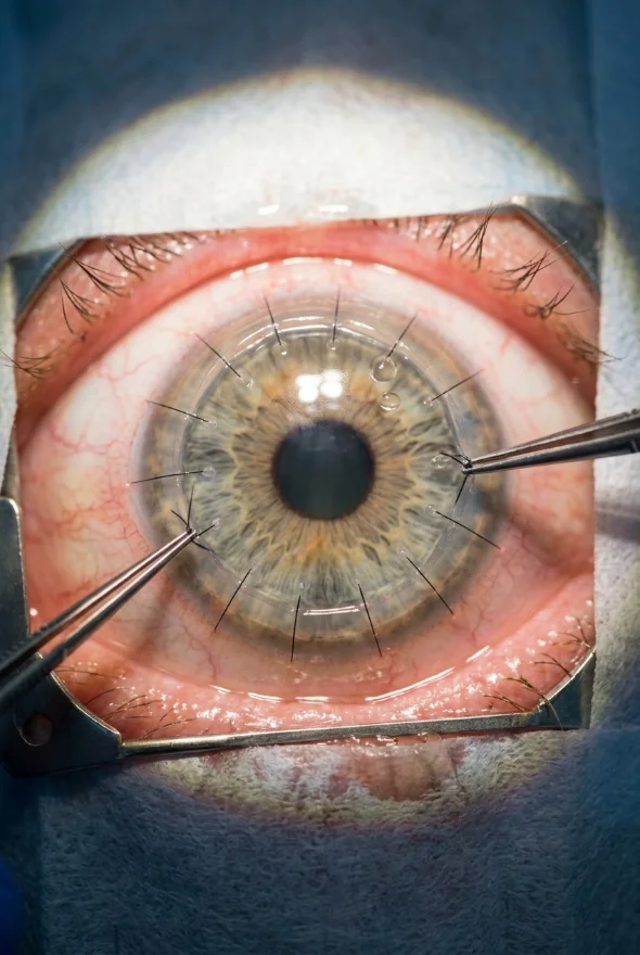
The corneal transplant success rate is high when the eye is otherwise healthy. For keratoconus, a 2023 study of 502 eyes found that 97.0% of grafts after DALK and 94.9% after full-thickness transplant (PK) were still working 20 years later. Rejection, the main risk, is least likely after partial-thickness surgery. Vision recovers in stages over 6 months to 2 years, depending on the technique. At Laser Istanbul Clinic, cornea specialist Doç. Dr. (Associate Professor) Muhammet Derda Özer plans and follows your transplant.
What is the corneal transplant success rate?
Most corneal transplants succeed, but "success" means two things: the graft keeps working (graft survival) and you see well enough to benefit (visual outcome). Studies report both, and they do not always move together. A clear graft with a lot of astigmatism can still need glasses or a contact lens, and a graft that sees well for years can still fail slowly.
A review in Clinical Ophthalmology (2022) gives these typical figures for the four main operations. They are ranges from published series, mostly in mixed groups of patients, so they are a guide and not a promise for your eye.
| PK (full thickness) | DALK (front and middle layers) | DSAEK (inner layer plus a thin layer of support tissue) | DMEK (inner layer only) | |
|---|---|---|---|---|
| What is replaced | The whole cornea, held with stitches | Front and middle layers; your own inner layer stays | The endothelium (inner pump layer) with some support tissue | The endothelium and its membrane only |
| Graft survival | 98% at 2 years; 90% at 5 years; 60–80% at 10 years (mixed diagnoses). In keratoconus: 98% at 2 years and 89% at 10 years | 93.9% at 10 years (mixed diagnoses); 90.0–90.4% at 5 years in keratoconus | 78.4–94.2% at 5 years (mixed diagnoses); 97% at 5 and 92% at 10 years in Fuchs' dystrophy | 90.0–97.4% at 5 years (mixed diagnoses) |
| Rejection rate | 21–31% | 0.8–10.9% | 2.2–7.9% | 0.1–5% |
| Speed of visual recovery | Slowest | Intermediate | Faster than PK | Fastest |
| Share of UK transplants, 2019–20 | 27.0% | 9.3% | 33.0% | 23.1% |
Source: Liu, Wong and Walkden, Clinical Ophthalmology 2022 (see Sources). DMEK became the most common transplant in Germany from 2016 and made up 65% of all keratoplasties there in 2021, up from 34% in 2013 (Flockerzi et al., British Journal of Ophthalmology 2024).
What this means for you: the technique is chosen for the disease, not for the headline number. Which corneal layer is diseased decides which operation is possible, and your surgeon should explain why one technique suits your eye better than another.
How successful is a corneal transplant for keratoconus?
Very successful: keratoconus is classed as a low-risk reason for a transplant. A 2019 review in Transplantation reports that first transplants in keratoconus survive 95% at 4 years and 92% at 5 years.
The best long-term evidence comes from a 2023 British Journal of Ophthalmology study of 502 keratoconus eyes (228 DALK, 274 PK) by Borderie and colleagues:
- Graft survival at 20 years: 97.0% after DALK and 94.9% after PK, with no statistically significant difference between them.
- Vision: on average from about 20/378 before surgery to about 20/32 at 30 months.
- Rejection: at least one rejection episode in 16.7% of DALK eyes and 19.0% of PK eyes.
The authors concluded that DALK, which keeps your own endothelium (the inner pump layer), may be preferable to PK when it is technically possible, because it protects endothelial cells and carries a lower risk of raised eye pressure. The inner-layer operations, DMEK and DSAEK, are used for diseases of the endothelium such as Fuchs' dystrophy, not for keratoconus.
A transplant is not the first step for most people with keratoconus. The NHS describes it as an option when glasses and lenses no longer help enough, or when the eye is painful. For the background, read our guide to keratoconus symptoms, stages and treatment options, and see why early cross-linking matters in children and teenagers.
How successful are corneal transplants for Fuchs' dystrophy and other endothelial problems?
Endothelial transplants (DMEK, DSAEK) restore vision faster and are rejected less often than PK, but the long-term picture is still being measured. Fuchs' endothelial dystrophy, where the cornea swells because its inner pump layer wears out, is the commonest reason for these operations (43% of German transplants in 2021).
Three studies show the range of results:
| Study | Patients | Result |
|---|---|---|
| Bichet et al., PLoS One 2023 (DMEK, one centre) | 107 eyes, 94% with Fuchs' dystrophy | 5-year graft survival 88%. At 5 years, 92% saw 20/40 or better, 77% saw 20/25 or better and 57% saw 20/20 or better. Rejection occurred in 1% of eyes. 18% needed at least one gas-bubble re-injection (rebubbling) for a partly detached graft. |
| van Rooij et al., PLoS One 2018 (Fuchs', Rotterdam, 2002–2013) | 721 first grafts: 171 PK, 91 posterior "mushroom" PK, 459 DSAEK | No significant difference in graft survival over 10 years. Rejection in 4.1% (PK) against 0.9% (DSAEK). Astigmatism at 2 years: 4.6 D after PK and 1.7 D after DSAEK. |
| Wilhelm et al., Scientific Reports 2025 (Fuchs', one German centre, first transplants 2003–2023) | 2,956 eyes: 2,672 DMEK, 204 PK, 80 DSAEK | 10-year graft survival 75% for DMEK, 73% for DSAEK and 92% for PK. 10-year rejection: 10% DMEK, 19% DSAEK, 13% PK. Median time to 6/12 vision: 7.8 months (DMEK), 12.4 months (DSAEK), 37.9 months (PK). |
Read the last row carefully. The authors found that endothelial grafts lose their cells faster over a decade, and they also warn that surgeons' and patients' willingness to repeat a full-thickness graft may skew the survival figures, because "failure" was defined as needing another transplant. The balanced message: DMEK gives the quickest vision recovery with a low rejection risk, and some patients will need a repeat procedure years later. None of these studies predicts the result for one particular eye.

How likely is rejection, and what are the warning signs?
Rejection means your immune system attacks the donor tissue. It is the most common cause of graft failure, it can often be treated if caught early, and it is less likely after partial-thickness surgery. The American Academy of Ophthalmology (AAO) says rejection is a problem for up to 3 in 10 people after a full-thickness transplant, and that prompt steroid treatment can sometimes reverse it. Partial-thickness operations carry a lower risk.
Use the table above as a guide: 21–31% for PK against 0.1–5% for DMEK in the 2022 review. Long-term cohorts give somewhat different numbers (10-year rejection of 10–19% in the 2025 German series), so surgeons talk in ranges and not in single figures.
Contact your surgeon the same day, or go to an eye emergency service, if you notice (NHS and AAO):
- a red eye
- eye pain
- sensitivity to light
- blurred or clouded vision
- sudden floaters, flashes or a dark shadow across your vision (the NHS lists these too; they can point to a retinal problem and need urgent review)
What makes a corneal transplant more likely to fail?
The main risk factors are abnormal blood vessels in the cornea, a previous graft, inflammation before surgery and high eye pressure. The 2019 Transplantation review describes "high-risk" recipients as those in whom rejection episodes occur in 30–60% of grafts and up to 70% fail within 10 years. It reports:
- Blood vessels growing into the cornea (neovascularisation): 4-year survival of a full-thickness graft falls from 83% with no vessels to 50% when all four quadrants are involved.
- Repeat transplants: 4-year survival of 86% for a second graft, 71% for a third and 56% for a fourth or later.
- Inflammation or steroid use before surgery: 85% against 58% survival at 4 years.
- Raised eye pressure or glaucoma: 81% against 55%.
If you have one of these factors, ask your surgeon how it changes the plan and the odds. A high-risk eye does not make a transplant impossible, but it calls for an experienced surgeon and closer follow-up.
How long is corneal transplant recovery?
Recovery has two clocks: the first two weeks, when the eye settles, and the following 6 to 24 months, when vision slowly sharpens. The NHS gives full visual recovery as up to 6 months when only the inner layer is replaced, up to 18 months when the outer and middle layers are replaced, and up to 2 years after a full-thickness transplant. The AAO says it can take a year or more to recover fully.
- Operation day
The operation takes 1–2 hours (NHS). It is done under local anaesthetic with eye drops and an optional sedative, or under general anaesthetic. Most people go home the same day; some stay 1–2 nights.
- First 1–2 weeks
Swelling, redness and discomfort for up to 2 weeks. Around 2 weeks off work (NHS). Vision is blurred. A first check in the first week.
- First month
Frequent drops as your surgeon prescribes. No eye rubbing. The NHS also advises avoiding contact sports, swimming and eye make-up in the early weeks; your surgeon gives you the exact period.
- 1–6 months
After DMEK, vision improves steadily; in the French 5-year DMEK series most of the gain came in the first 3 months. Stitches (PK, DALK) stay in for several months and are removed gradually (NHS).
- 6–18 months
Vision keeps improving, especially after DALK (NHS: up to 18 months). Your surgeon decides when your glasses or contact-lens prescription can be finalised.
- Up to 2 years
Full visual recovery after a full-thickness transplant (NHS). Reviews are usually every few months for 1–2 years, sometimes longer.
- Long term
Drops continue long term (NHS), and you stay under review because late rejection and graft ageing are possible.
Sources: NHS cornea transplant guidance; AAO; Wilhelm et al. 2025; Bichet et al. 2023. Your surgeon gives you personal dates and your exact drop schedule.
How long until I can see clearly after a corneal transplant?
It depends on the technique. In the 2025 German Fuchs' study, the median time to reach 6/12 vision was 7.8 months after DMEK, 12.4 months after DSAEK and 37.9 months after PK. After keratoconus transplants in the French study, the average vision of about 20/32 was measured at 30 months. Do not expect your final sharp image at 3 months; ask your surgeon when a new prescription makes sense.
When can I drive, work and exercise?
The NHS advises around 2 weeks off work. Drive again only when your vision is good enough and your surgeon agrees. Avoid rubbing the eye, contact sports, swimming and eye make-up in the early weeks, as your surgeon directs.

What are the risks and complications of a corneal transplant?
The NHS lists these possible complications: transplant rejection, clouding of the cornea, the graft moving out of position (dislodgement), astigmatism, inflammation, raised eye pressure (glaucoma), infection, retinal detachment and cataract. The AAO adds bleeding. Several can be spotted early at routine reviews, which is why follow-up matters as much as the operation itself.
- Graft detachment after DMEK is the commonest technical issue and is usually fixed with a gas bubble (rebubbling). It affected 18% of eyes in the French 5-year DMEK series.
- Astigmatism is a main reason vision after full-thickness transplant is not sharp without correction: 4.6 D at 2 years after PK against 1.7 D after DSAEK in the Rotterdam series.
- Endothelial cell loss is expected. In the 5-year DMEK series, cell density fell by 65%, with nearly half of that loss in the first 6 months.
No surgeon can guarantee an outcome. A good surgeon tells you the realistic range for your eye before you decide.
What happens if a corneal transplant fails?
A failed graft can often be replaced, but a second transplant is less likely to last than the first. Repeat grafts survive less well (see the 86% and 71% figures above). In a 2023 series from Moorfields Eye Hospital in London of 104 eyes that needed a second transplant after a failed PK, 12-month graft survival was 93.4% for a repeat PK, 81.0% for DSAEK and 63.6% for DMEK; at 24 months it was 79.4%, 69.4% and 58.7%. The authors stress that careful patient selection by experienced surgeons is needed to choose the procedure.
Early review gives the best chance of saving a graft that is in trouble, which is why the warning signs above matter.
Having a corneal transplant in Istanbul: how follow-up works
Corneal transplants need longer follow-up than most eye operations, so plan it before you book. At Laser Istanbul Clinic, corneal disease, keratoconus and corneal transplant are the specialty of Doç. Dr. (Associate Professor) Muhammet Derda Özer, who speaks Turkish and English. The usual steps are:
Send your scans
Upload your prescription, corneal topography and any previous eye reports in the free online consultation. The surgeon tells you whether a transplant is the right next step and which technique he would consider.
Pre-operative examination in Istanbul
Your eye is examined and scanned again before surgery, with a personal interpreter and a coordinator beside you.
Surgery
by the named surgeon in a fully equipped, Ministry of Health–licensed operating theatre in Istanbul.
Follow-up checks in Istanbul
Your surgeon examines the eye before you travel; plan {{nights in Istanbul for a corneal transplant}} in the city. Exact dates are set once your scans have been reviewed. Hotel and airport transfers are arranged separately on request.
Follow-up at home
Your coordinator and surgeon follow your recovery remotely by video at agreed intervals. We help you arrange in-person checks, including stitch removal if needed, with a local ophthalmologist and share your full surgical records.
Because follow-up lasts 1–2 years or longer (NHS), the most important thing before you travel is to know who will examine your eye at home and how to reach them in an emergency.
Frequently asked questions
It depends on the technique and on why the transplant is needed. In low-risk eyes such as keratoconus, first grafts survive about 92% at 5 years (2019 review) and 94.9–97.0% at 20 years (2023 French study). In mixed groups, DMEK survives 90.0–97.4% at 5 years and PK about 90%. Graft survival is lower in high-risk eyes.
Most corneal transplants work and improve vision, particularly for keratoconus and for Fuchs' dystrophy treated with DMEK. In 107 DMEK eyes followed for 5 years, 92% saw 20/40 or better. Some grafts fail, mainly from rejection or gradual loss of endothelial cells, and a repeat transplant is sometimes needed.
Very successful. In 502 keratoconus eyes, 97.0% of DALK grafts and 94.9% of PK grafts were still working at 20 years, and average vision improved from about 20/378 to about 20/32. Many patients still need glasses or a contact lens for the sharpest vision.
About 2 weeks off work, then months of gradual improvement. The NHS gives up to 6 months for inner-layer transplants, up to 18 months for outer and middle layers, and up to 2 years for a full-thickness transplant. Follow-up reviews usually continue for 1–2 years or longer.
The operation is done under local or general anaesthetic. Afterwards the eye is usually uncomfortable, red and swollen for up to 2 weeks (NHS), which your surgeon manages with drops and any pain relief they advise. Severe or worsening pain needs a same-day review, because pain is one of the warning signs of rejection.
A red eye, eye pain, light sensitivity or blurred vision, as listed by the NHS and AAO. Sudden floaters, flashes or a dark shadow can point to a retinal problem. Contact your surgeon the same day: rejection caught early can often be reversed with steroid treatment.
Yes, but results are less predictable. Published 4-year survival is 86% for a second graft, 71% for a third and 56% for a fourth or later. Your surgeon assesses the cause of the first failure before choosing a technique.
Often preferable when it is possible, because it replaces only the diseased layer, recovers faster and carries a lower rejection risk. PK is still needed when all layers are affected. The right choice depends on which layer is diseased, so the surgeon decides after scans.
Thinking about a corneal transplant?
Doç. Dr. Muhammet Derda Özer reviews your corneal scans, tells you honestly whether a transplant is needed or whether other treatments are still enough, and explains which technique he would consider and what recovery to expect. Read our overview of keratoconus treatment in Turkey for the full range of options.
Sources for the facts on this page
- All opened and checked on 7 Oct 2026.
- NHS. Cornea transplant. https://www.nhs.uk/conditions/cornea-transplant/
- American Academy of Ophthalmology. Corneal transplant (reasons, types, rejection, recovery). https://www.aao.org/eye-health/treatments/corneal-transplant
- Liu S, Wong YL, Walkden A. Current Perspectives on Corneal Transplantation. Clinical Ophthalmology 2022. https://pmc.ncbi.nlm.nih.gov/articles/PMC8904759/
- Borderie VM, et al. Long-term outcomes of deep anterior lamellar versus penetrating keratoplasty for keratoconus. British Journal of Ophthalmology 2023;108(1):10–16. https://pmc.ncbi.nlm.nih.gov/articles/PMC10803977/
- Armitage WJ, Goodchild C, Griffin MD, et al. High-risk Corneal Transplantation: Recent Developments and Future Possibilities. Transplantation 2019. https://pmc.ncbi.nlm.nih.gov/articles/PMC6867666/
- Wilhelm TI, Gauché L, Böhringer D, et al. Ten-year outcomes after DMEK, DSAEK, and PK. Scientific Reports 2025. https://pmc.ncbi.nlm.nih.gov/articles/PMC11707042/ (DOI 10.1038/s41598-025-85138-4)
- Bichet P, Moskwa R, Goetz C, et al. Five-year clinical outcomes of 107 consecutive DMEK surgeries. PLoS One 2023. https://pmc.ncbi.nlm.nih.gov/articles/PMC10735023/
- van Rooij J, Lucas EH, Geerards AJ, et al. Corneal transplantation for Fuchs' endothelial dystrophy: a comparison of three surgical techniques. PLoS One 2018. https://pmc.ncbi.nlm.nih.gov/articles/PMC6173378/
- Roberts HW, de Benito-Llopis L. Comparison of repeat penetrating keratoplasty, DSAEK and DMEK. Eye 2023. https://pmc.ncbi.nlm.nih.gov/articles/PMC10686404/
- Flockerzi E, Turner C, Seitz B, et al. Descemet's membrane endothelial keratoplasty is the predominant keratoplasty procedure in Germany since 2016. British Journal of Ophthalmology 2024. https://pmc.ncbi.nlm.nih.gov/articles/PMC11137454/