
- Home
- Knowledge centre
- Keratoconus & cornea guides
- Stages of Keratoconus: Symptoms, Causes and Treatment Options
Stages of Keratoconus: Symptoms, Causes and Treatment Options
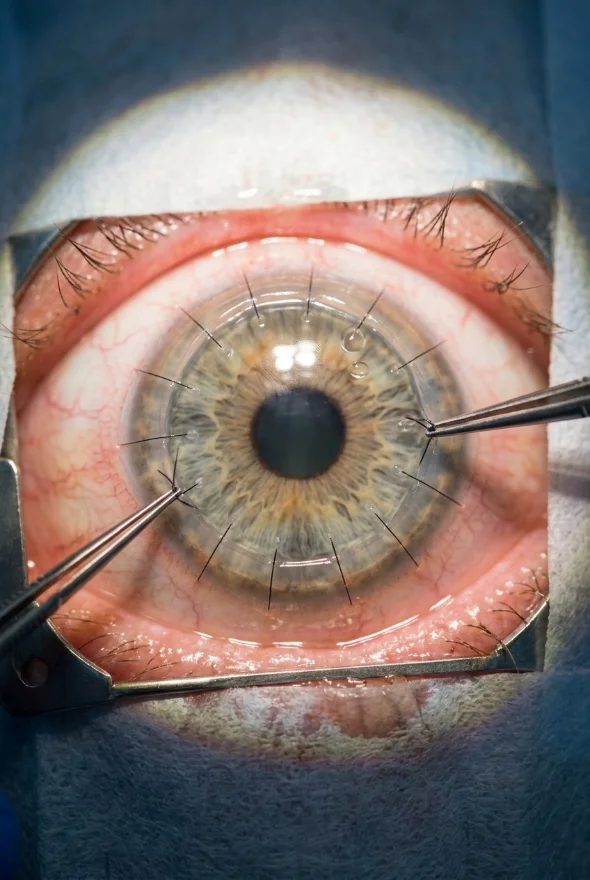
The stages of keratoconus run from stage 1 (an early, mild cone) to stage 4 (a very thin, scarred cornea), most often graded with the Amsler-Krumeich system. Symptoms begin with blurred, distorted vision and a prescription that keeps changing. Treatment depends on whether the cornea is getting worse: cross-linking stops progression, while lenses, corneal rings or a transplant improve vision. Doç. Dr. Muhammet Derda Özer ("Doç." means Associate Professor) leads keratoconus care at Laser Istanbul Clinic.
What is keratoconus?
Keratoconus is an eye condition in which the cornea, the clear dome at the front of the eye, gradually thins and bulges forward into a cone shape. The American Academy of Ophthalmology (AAO) describes it as the cornea thinning out and bulging like a cone. Because the cornea does most of the eye's focusing, a cone bends light unevenly and vision becomes blurred and distorted in a way that glasses correct less and less well.
It often starts in the late teens or early twenties, and the AAO says vision symptoms slowly get worse over about 10 to 20 years, although the pace varies a great deal from person to person. NHS eye-hospital information stresses that keratoconus does not cause blindness and that, even in advanced stages, vision can usually still be corrected with contact lenses.
It is also more common than older textbooks suggest. EyeWiki, the AAO's clinical encyclopaedia, quotes a prevalence of about 1 in 700 and notes higher rates in the Middle East, India, China and Australia. A nationwide Dutch registry study of 4.4 million people, published in 2017, found a prevalence of about 1 in 375, with an average age at diagnosis of 28.3 years and 60.6% of patients male.
What causes keratoconus?
The exact cause of keratoconus is not known. The AAO lists genetics, eye allergies, excessive eye rubbing and connective-tissue disorders such as Marfan and Ehlers-Danlos syndromes as associated factors, and NHS eye-hospital information says both genetic and environmental factors play a part. In other words, keratoconus is not one single cause but a weak cornea meeting a trigger.
The keratoconus causes and risk factors that doctors describe most often are:
- Family history. About 1 in 10 people with keratoconus have a parent who has it too (AAO), but it is not usually classed as an inherited disease. Brothers, sisters and children of someone with keratoconus are worth screening.
- Eye rubbing. EyeWiki lists eye rubbing, often linked to eye allergy, as a modifiable risk factor. The AAO warns that rubbing can damage thin corneal tissue and make symptoms worse.
- Allergic disease. Atopy (hay fever, eczema, asthma) and allergic eye disease, which make people rub their eyes. NHS eye-hospital information says keratoconus is seen more often in people with asthma and eczema.
- Some general conditions. EyeWiki lists Down syndrome, connective-tissue disorders, sleep apnoea, hypothyroidism, Leber congenital amaurosis and retinitis pigmentosa, and hormonal change in pregnancy.
At the level of the tissue, EyeWiki describes breaks in, or absence of, the cornea's Bowman layer, disorganised collagen, scarring and thinning, with raised inflammatory chemicals in the tear film. A weakened cornea can no longer hold its dome shape.
What causes keratoconus to get worse?
Keratoconus gets worse mainly in younger people and when the cornea is mechanically stressed, for example by eye rubbing. Because the cornea naturally stiffens with age, NHS eye-hospital information says keratoconus usually stops getting worse by the mid-30s, and a review in Community Eye Health (2024) calls young age the most important risk factor for progression.
| What can make it progress | What the evidence says |
|---|---|
| Young age | Children present with more advanced and more rapidly progressive disease than adults (EyeWiki). In a 2020 study of 41 children and teenagers by Doç. Dr. Özer's team, age at presentation was the only factor that predicted progression 2 years after cross-linking: 35% of those aged 14 or under progressed, against 4% of those aged 15 to 18. |
| Eye rubbing | Named by the AAO and EyeWiki as a risk factor that can worsen the condition. It also raises the risk of a sudden complication called hydrops (see below). |
| Allergy and itchy eyes | Allergic eye disease drives rubbing. In children, a history of allergy was linked to faster progression (EyeWiki). |
| A steeper cone at diagnosis | In children, a maximum corneal curvature (Kmax) of 55 D or more at the start predicted faster progression (EyeWiki). |
| Pregnancy | Hormonal changes in pregnancy are named as a possible trigger for progression (Community Eye Health, 2024; EyeWiki). |
| Sleep apnoea | Listed by EyeWiki as a modifiable risk factor. |
| Time without follow-up | Progression may continue beyond the third decade, and even after treatment, so long-term follow-up matters (EyeWiki). |
What you can do: the one change fully in your control is to stop rubbing your eyes. If itching drives it, ask your optometrist or doctor about treating the allergy. Nothing in this table means rubbing, allergy or pregnancy "caused" your keratoconus: they are factors that can tip a susceptible cornea.
What are the symptoms of keratoconus?
Keratoconus symptoms start subtly and are often mistaken for ordinary astigmatism or short sight. The AAO lists these signs:
- Early stage: mild blurring of vision, distorted vision in which straight lines look bent or wavy, more sensitivity to light and glare, and sometimes redness or swelling of the eye.
- Later stage: more severe blurring and distortion, increasing short-sightedness and astigmatism that need frequent prescription changes, and contact lenses that no longer fit or feel comfortable.
NHS eye-hospital information puts the classic warning sign simply: worsening vision that causes frequent changes to your prescription. Light sensitivity and glare are also typical. EyeWiki describes the main symptom as progressive changes in vision that are not easily corrected with glasses.
An eye specialist may also see signs you cannot: a conical reflection on the cornea, fine vertical lines called Vogt striae, an iron-coloured ring at the base of the cone (the Fleischer ring), and, late in the disease, a bulge of the lower eyelid when you look down (Munson sign), all described by EyeWiki. Only a corneal scan can confirm the diagnosis.
Sudden pain or sudden loss of vision: acute hydrops
Occasionally the thin cornea splits on its inside surface (the Descemet membrane) and fluid floods into the cornea. EyeWiki describes this as acute corneal hydrops: sudden loss of vision, light sensitivity and discomfort, sometimes triggered by eye rubbing. It affects about 0.2% to 2.8% of people with keratoconus, most often people aged 20 to 40. Many cases settle by themselves within 2 to 4 months, but the cornea may scar, so a sudden drop in vision should be seen by an eye specialist promptly.
What are the stages of keratoconus?
The stages of keratoconus describe how far the cornea has changed, from stage 1 (mild) to stage 4 (advanced). The classic grading is the Amsler-Krumeich classification, which uses the induced short sight and astigmatism, the average corneal curvature (K reading), the thickness of the thinnest point and any scarring.
| Stage | Prescription (myopia and/or astigmatism) | Mean corneal curvature (K) | Thinnest cornea | Scarring |
|---|---|---|---|---|
| Stage 1 | under 5 D | 48 D or less; Vogt striae and/or eccentric steepening | not specified | none |
| Stage 2 | 5–8 D | under 53 D | over 400 µm | none |
| Stage 3 | 8–10 D | over 53 D | 200–400 µm | none |
| Stage 4 | not measurable | over 55 D | under 200 µm | central scar |
Criteria as set out by Khorrami-Nejad et al., 2019. A "D" is a dioptre, the unit of lens power; 1 µm (micrometre) is a thousandth of a millimetre.
What do stages 1, 2, 3 and 4 mean in practice?
- Stage 1 (early): you may see well with glasses or soft lenses; a scan shows a mild cone, and the main symptoms are slight blur, ghosting and glare.
- Stage 2 (moderate): the cone is steeper and vision is distorted in glasses; rigid contact lenses often help much more. The cornea is still thicker than 400 µm, which is the usual minimum thickness for standard cross-linking (EyeWiki).
- Stage 3 (advanced): a much thinner, steeper cornea; glasses give little help and contact lenses may be hard to fit.
- Stage 4 (severe): a central scar and a cornea thinner than 200 µm; vision is poor even with lenses, and a transplant is usually considered.
Newer grading: the ABCD system
The Amsler-Krumeich system looks only at the front surface of the cornea, so it can miss early disease. The ABCD classification, published by Belin and colleagues in 2020, adds the back surface and your best corrected vision, scoring four things: A, the curvature of the front surface; B, the curvature of the back surface; C, the thickness at the thinnest point; and D, your best-corrected distance vision.
| ABCD stage | Thinnest point (C) | Best-corrected vision (D) |
|---|---|---|
| 0 | over 490 µm | 20/20 |
| I | over 450 µm | worse than 20/20 |
| II | over 400 µm | worse than 20/40 |
| III | over 300 µm | worse than 20/100 |
| IV | 300 µm or less | worse than 20/400 |
Thresholds for the front-surface radius (A) and back-surface radius (B) are also part of each stage; see Belin et al., 2020.
An important point: a stage is a snapshot of the cornea's shape on one day. It does not tell you how fast you are changing, and it does not by itself decide your treatment. A young person with stage 1 who is progressing may need cross-linking urgently; a 40-year-old with a stable stage 3 may need only lenses.
How do doctors know keratoconus is getting worse?
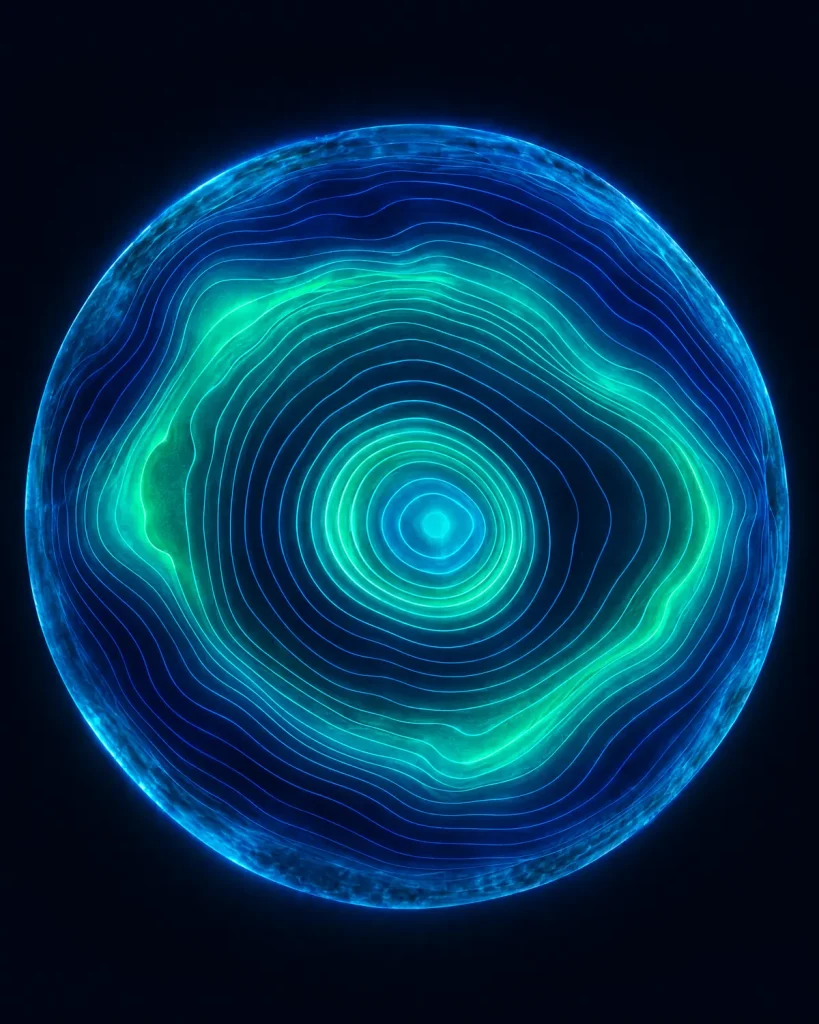
Doctors confirm keratoconus and measure progression with corneal imaging, repeated over time. NHS eye-hospital information describes diagnosis by eye examination, slit-lamp examination and corneal topography, a quick, painless picture that checks the shape and thickness of the cornea, with several follow-up visits over years to track change. The Community Eye Health review adds scans that map both the front and back of the cornea and OCT scans that show thinning.
Progression is a measured change, not a feeling. In one Australian trial described by EyeWiki, progression over 12 months meant an increase of 1 D or more in astigmatism, an increase of 1 D or more in the steepest corneal reading (Kmax), or a flattening of the best-fitting contact-lens curve by more than 0.1 mm. EyeWiki also suggests reviewing children about every 3 to 6 months, sooner when progression is suspected.
What are the treatment options for keratoconus, stage by stage?
Keratoconus treatment has two separate goals: stop the cornea getting worse (cross-linking) and improve vision (lenses, corneal rings, an implantable lens or a transplant). Many patients need one from each group.
| Stage | Usual options | Notes |
|---|---|---|
| Stage 1 | Glasses or soft contact lenses; cross-linking if scans show progression | The AAO lists glasses or soft lenses for the early stages. |
| Stage 2 | Rigid contact lenses; cross-linking if progressing; corneal rings if lenses are not tolerated | Rigid lenses replace the uneven cone with a smooth optical surface; most people who can wear them see a dramatic improvement (EyeWiki). |
| Stage 3 | Rigid, hybrid or scleral lenses; cross-linking if the cornea is thick enough; corneal rings or surgery in selected eyes | Thin corneas (under 400 µm) can sometimes be treated with modified protocols (EyeWiki). |
| Stage 4 | Corneal transplant when lenses no longer give useful vision | The NHS says a transplant is considered when glasses or lenses are no longer enough. |
A general guide only. Your surgeon recommends a plan after examining your eyes; the stage alone never decides it.
Corneal cross-linking: the treatment that can stop keratoconus
Corneal cross-linking (CXL) stiffens the cornea with riboflavin (vitamin B2) drops activated by ultraviolet-A light, and it is the main treatment that targets progression itself. NHS eye-hospital information reports success in halting progression in more than 90% of treated eyes, with that hospital's own data since 2013 at about 94% and fewer than 1% needing repeat treatment. An NHS trust leaflet quotes a lower figure, "more than seven people out of every 10", so success rates differ between centres and between ways of measuring it.
Cross-linking aims to stabilise the cornea, not to improve vision, so you will usually still need glasses or lenses afterwards. Recovery is a few uncomfortable days (an NHS trust leaflet says 2 to 3 days of pain and light sensitivity), at least one week off work, and drops for about four weeks. The US Food and Drug Administration approved cross-linking on 18 April 2016, and in October 2025 approved the first epithelium-on system, which leaves the surface skin of the cornea in place (EyeWiki). Risks include temporary haze, infection and scarring; serious problems are uncommon but real.
Because cross-linking works best before the cornea has thinned too far, it is usually offered when progression is documented, most often in younger people. NHS eye-hospital information says it is generally not needed once you are past your mid-to-late 30s, because the cornea stiffens naturally.
Our cornea specialist, Doç. Dr. Muhammet Derda Özer, has published studies on accelerated cross-linking in children and teenagers. If you are considering treatment abroad, read about keratoconus treatment in Turkey.
Corneal rings, an implantable lens or laser?
Corneal rings are two small curved segments placed in the outer cornea to flatten the cone; a 2016 review in Eye and Vision reports an average flattening of 3–5 D, and rings do not stop progression on their own, so they are often combined with cross-linking. Standard laser eye surgery (LASIK) is not suitable for keratoconus: the US Food and Drug Administration lists keratoconus and thin corneas as reasons not to have it. Once the cornea is stable, other options such as an implantable lens can be considered.
Corneal transplant
A transplant is for advanced disease. According to an NHS trust leaflet, about 20 in 100 untreated patients may eventually need one; a Dutch national study found about 25% fewer transplants for keratoconus in the three years after cross-linking was introduced (201 against 269). NHS information says the operation takes 1 to 2 hours, most people go home the same day, around 2 weeks off work is advised, and full visual recovery takes up to 18 months when the outer and middle layers are replaced and up to 2 years for a full-thickness transplant.

Will keratoconus make me blind?
No. NHS eye-hospital information says keratoconus does not cause blindness, and that vision can usually be corrected with contact lenses even when it is advanced. The aim of monitoring and cross-linking is to protect your useful vision and avoid a transplant. Poorly controlled disease can seriously limit reading, driving and work, which is why early diagnosis matters.
When should you see a doctor?
See an optometrist or eye doctor soon if your glasses prescription keeps changing, your vision stays blurred or distorted even with new glasses, lights streak or ghost, or contact lenses stop fitting. Ask for corneal topography or tomography; a basic sight test alone can miss early keratoconus.
Get an urgent eye check if you have sudden loss of vision, new severe pain or a rapidly clouding eye, because this can be hydrops or an infection.
Book a review if you already have keratoconus and have not had a scan for a year or more, you are pregnant, or you are a teenager or young adult with diagnosed keratoconus: this is the age when progression is fastest.
Frequently asked questions
Keratoconus is a condition in which the cornea thins and bulges into a cone shape, causing blurred, distorted vision and light sensitivity. It usually begins in the teens or twenties and can change over 10 to 20 years. It does not cause blindness and usually remains correctable with lenses.
The exact cause is unknown. Genetics, eye allergies, eye rubbing and connective-tissue disorders are all linked to it, and about 1 in 10 people with keratoconus have an affected parent.
Young age is the strongest factor, together with eye rubbing, allergic eye disease, a steeper cornea at diagnosis, and possibly pregnancy. In children, progression is faster. In adults past the mid-30s, keratoconus usually stops progressing as the cornea stiffens with age.
Typical keratoconus symptoms are blurred or distorted vision, straight lines looking wavy, glare and light sensitivity, and a glasses prescription that keeps changing. Later, contact lenses fit poorly and astigmatism rises. Sudden pain or a sudden loss of vision needs urgent attention.
Most doctors use four stages (Amsler-Krumeich stages 1 to 4), from mild to severe. A newer system, the Belin ABCD classification, uses stages 0 to IV and also scores the back surface of the cornea and your best-corrected vision.
Stage 2 is moderate: a prescription of about 5–8 D, a cornea still thicker than 400 µm and no scarring. It is a good time to act if scans show progression, because the cornea is thick enough for standard cross-linking.
No. Treatment cannot restore a normal cornea, but cross-linking can stabilise it, and lenses, rings or surgery can improve how well you see.
Sometimes. Around 1 in 10 people with keratoconus have an affected parent, but it is not usually considered a classically inherited disease, and many cases occur with no family history.
Yes. Send your corneal scans and prescription for a free review; your surgeon can tell you from the scans whether your keratoconus appears to be progressing and which treatment may suit your eyes.
Worried your keratoconus is progressing?
Doç. Dr. Muhammet Derda Özer, our cornea and keratoconus specialist, reviews your scans, explains your stage in plain language and recommends the right next step for your eyes. Prof. Dr. Ahmet Satıcı and Op. Dr. Aysan Bayram join the plan when laser, lens or cataract decisions arise once the cornea is stable.
Sources for the facts on this page
- American Academy of Ophthalmology. What Is Keratoconus? https://www.aao.org/eye-health/diseases/what-is-keratoconus
- EyeWiki (AAO). Keratoconus. https://eyewiki.org/Keratoconus
- EyeWiki (AAO). Corneal Collagen Cross-Linking. https://eyewiki.org/Corneal_Collagen_Cross-Linking
- EyeWiki (AAO). Acute Corneal Hydrops. https://eyewiki.org/Acute_Corneal_Hydrops
- NHS eye-hospital patient information. Keratoconus. https://www.moorfields.nhs.uk/eye-conditions/keratoconus
- NHS eye-hospital patient information. Keratoconus: diagnosis and treatment. https://www.moorfields.nhs.uk/eye-conditions/keratoconus/diagnosis-and-treatment
- NHS eye-hospital patient information. Corneal cross-linking. https://www.moorfields.nhs.uk/eye-conditions/keratoconus/corneal-cross-linking
- NHS trust patient leaflet. Corneal cross-linking (CXL). https://www.dgft.nhs.uk/pil/corneal-cross-linking-cxl/
- NHS. Cornea transplant. https://www.nhs.uk/conditions/cornea-transplant/
- Khorrami-Nejad M, et al. Changes in Corneal Asphericity after MyoRing Implantation in Moderate and Severe Keratoconus. J Ophthalmic Vis Res 2019 (Amsler-Krumeich criteria). https://pmc.ncbi.nlm.nih.gov/articles/PMC6825702/
- Belin MW, et al. ABCD: A new classification for keratoconus. Indian J Ophthalmol 2020. https://pmc.ncbi.nlm.nih.gov/articles/PMC7856970/
- Godefrooij DA, et al. Age-specific Incidence and Prevalence of Keratoconus: A Nationwide Registration Study. Am J Ophthalmol 2017 (PMID 28039037). https://pubmed.ncbi.nlm.nih.gov/28039037/
- Godefrooij DA, et al. Nationwide reduction in the number of corneal transplantations for keratoconus following the implementation of cross-linking. Acta Ophthalmol 2016 (PMID 27213687). https://pubmed.ncbi.nlm.nih.gov/27213687/
- Deshmukh R, Gore D. Keratoconus: diagnosis and management. Community Eye Health 2024. https://pmc.ncbi.nlm.nih.gov/articles/PMC11921258/
- Ozer MD, et al. Comparison of the efficacy of accelerated corneal cross-linking therapy in different pediatric age groups having progressive keratoconus. Int Ophthalmol 2020 (PMID 32488590). https://pubmed.ncbi.nlm.nih.gov/32488590/
- Vega-Estrada A, Alió JL. The use of intracorneal ring segments in keratoconus. Eye and Vision 2016. https://pmc.ncbi.nlm.nih.gov/articles/PMC4791885/
- US Food and Drug Administration. When is LASIK not for me? https://www.fda.gov/medical-devices/lasik/when-lasik-not-me