
Corneal Topography: The Map of Your Cornea Before Eye Surgery
Corneal topography is a quick, painless scan that maps the shape of your cornea, the clear window at the front of your eye. It is the first test your surgeon looks at before laser eye surgery, the key test for finding and following keratoconus, and an important part of planning lens and cataract surgery. At Laser Istanbul Clinic it is part of every pre-operative examination, and your surgeon reads the result with you.

Corneal topography is a no-touch scan that maps the curvature of your cornea like a contour map of hills and valleys. Each scan takes a few seconds. It shows astigmatism, early keratoconus and irregular corneas that would make laser eye surgery unsafe, and it guides the laser and lens-power plan. Abnormal topography is the single most significant known risk factor for weakening of the cornea after LASIK.
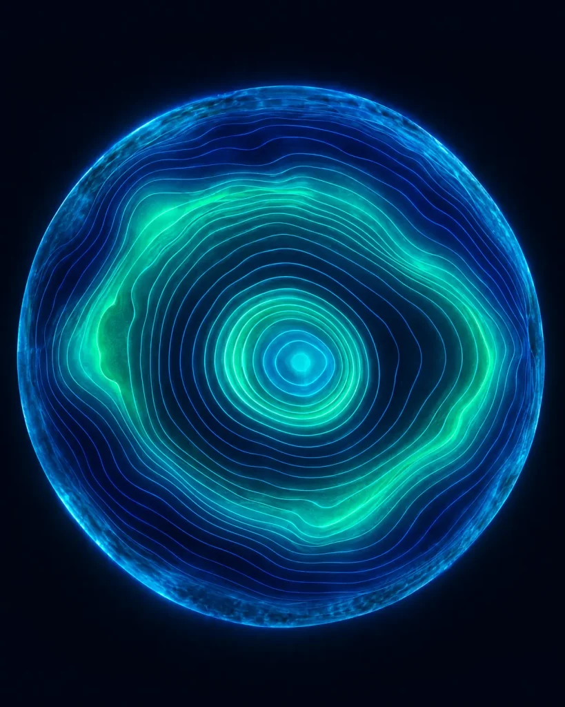
What corneal topography measures
A contour map of the front of your eye
Your cornea provides about two-thirds of your eye's focusing power, so tiny differences in its shape change how clearly you see. A normal cornea is smooth and slightly flatter towards the edge. Corneal topography measures that shape point by point and turns it into colour maps.
- Curvature and power of the front surface, in dioptres, across the whole cornea, not just the centre
- Astigmatism: how much, at what angle, and whether it is regular (symmetrical) or irregular
- Elevation: areas that sit higher or lower than a perfect reference shape
- Thickness (pachymetry) at every point, including the thinnest point, on tomography devices
- The back surface of the cornea, also on tomography devices, where early keratoconus often shows first
Topography or tomography? Placido or Pentacam?
You will see both words. Strictly, topography maps the front surface, while tomography builds a three-dimensional picture of the front surface, back surface and thickness. EyeWiki notes that topography is good at capturing the power of the front of the cornea, while the overall shape is best captured by tomography.
| Corneal topography machine type | How it works | What it measures | Best for |
|---|---|---|---|
| Placido-disc topographer | Projects illuminated concentric rings onto the eye and photographs their reflection from the tear film | Front-surface curvature and power | Astigmatism, contact-lens fitting, surface regularity |
| Rotating Scheimpflug camera (e.g. Pentacam) | A rotating camera and slit of blue light capture up to 25 slit images in under two seconds, with up to 25,000 data points | Front and back surfaces, thickness map, 3D model of the front of the eye | Screening for early keratoconus and laser safety |
| Scanning-slit and anterior-segment OCT systems | Light slits or optical coherence tomography scan across the cornea | Front and back surfaces and thickness | Alternative tomography method |
In practice
So "corneal topography Pentacam" usually means a Scheimpflug tomography scan. According to EyeWiki, rotating Scheimpflug imaging currently gives the most useful information for spotting early weakening (ectasia) of the cornea.
Our examination uses {{topographer / tomographer models used by your surgeons}}.
Why corneal topography is essential before laser eye surgery
Laser eye surgery removes a precise amount of corneal tissue. That is safe in a healthy cornea, and it can be harmful in a cornea that is already weak. The test exists to tell the two apart before any laser is used.
1. It screens out corneas at risk of ectasia
The most feared complication of LASIK is ectasia, when a weakened cornea gradually bulges and vision distorts. EyeWiki describes abnormal corneal topography or tomography as "the single most significant identifiable risk factor" for ectasia after LASIK. It can reveal keratoconus and forme fruste (hidden) keratoconus that glasses tests and a normal eye examination miss. EyeWiki reports that Scheimpflug imaging and screening software have reduced ectasia to roughly 1 in 5,000 cases.
2. It decides which laser treatment is safe
Together with corneal thickness, the scan shows how much tissue can be removed while leaving enough untouched cornea (at least 250 microns under a LASIK flap, with 300 preferred by many surgeons). If the margin is too small, your surgeon may suggest a surface treatment, SMILE Pro or an implantable lens instead of LASIK.
3. It plans the treatment itself
The American Academy of Ophthalmology explains that topography helps the surgeon understand exactly how to reshape the cornea during refractive surgery.
4. It checks that your cornea is stable
Contact lenses, especially rigid and overnight orthokeratology lenses, temporarily change the corneal shape. EyeWiki advises that topography should be stable on two separate visits before final planning when lens-induced changes are suspected.
Before lens and cataract surgery, topography helps choose the right lens. It shows whether astigmatism is regular enough for a toric lens, whether the cornea suits a multifocal lens, and, in people who have had laser surgery in the past, gives a more accurate picture of corneal power than standard keratometry.
The Royal College of Ophthalmologists also advises that anyone who has had laser vision correction tell their cataract surgeon, because it changes the lens-power calculation.
Corneal topography and keratoconus
The key test for finding and following keratoconus
Keratoconus is a condition in which the cornea gradually thins and bulges into a cone, causing blurred, distorted vision and irregular astigmatism. EyeWiki notes that its prevalence is often reported as 1 in 700, with considerably higher rates in the Middle East, India, China and Australia, and that it usually begins in adolescence and can progress into the mid-20s and 30s.
Diagnosis
A review in Community Eye Health (2024) states that a definitive diagnosis of keratoconus is made with corneal topography and/or tomography. Typical signs on the maps include steepening that is stronger in the lower half of the cornea (inferior steepening), an asymmetric "bow-tie" pattern or a skewed axis, and thinning and raised elevation in the same area, often seen first on the back surface.
Monitoring
Keratoconus is followed with repeated scans, ideally on the same device. The same review suggests three consecutive scans per eye at each visit and monitoring every 6–12 months, or every 3 months in high-risk patients. An increase in corneal curvature of more than 1 dioptre, or thinning of more than 15 microns, may be considered significant.
Deciding on treatment
Corneal cross-linking strengthens the cornea to stop keratoconus getting worse. A Leeds Teaching Hospitals NHS Trust leaflet explains that it is most suitable for progressive keratoconus, including patients whose corneal topography maps show clear progression, and that it prevents further vision loss in about 95% of patients. Topography also guides corneal ring segments, specialist contact lenses and, in advanced cases, corneal transplantation.

What to expect
The corneal topography procedure
Before the test
- Take a break from contact lenses, so your cornea returns to its natural shape. Typical advice is at least 1 week for soft lenses and 2 weeks or more for rigid lenses. Before a laser eye surgery assessment, the US FDA advises 2 weeks for soft lenses, 3 weeks for toric soft or rigid gas-permeable lenses and 4 weeks for hard lenses. Your coordinator will confirm the timing for your lenses.
- Bring your glasses, your current prescription and any previous corneal scans. Earlier scans are especially valuable for keratoconus, because they show change over time.
During the test
- You sit facing the machine with your chin and forehead resting on a support.
- You look at a fixed light or target inside the device and keep your eye open.
- The device captures the images. Nothing touches your eye, and there is no pain.
- The scan is usually repeated a few times for each eye to make sure the readings are consistent. A dry or uneven tear film can blur the map, so you may be asked to blink and try again.
How long does it take? The American Academy of Ophthalmology notes that each scan takes only a few seconds, though it may need to be repeated a few times. Corneal topography is one part of your full pre-operative examination, which also includes your prescription, eye pressure and a retina check.
After the test: topography itself has no after-effects. Your surgeon goes through the maps with you at the same appointment.
How corneal topography results are read
Corneal topography interpretation is a specialist skill. Your surgeon looks at the patterns, the numbers and the two eyes side by side, together with your prescription and examination findings. These are the main elements.
Colour scales
Warm colours (red and orange) show steeper or higher areas; cool colours (blue and purple) show flatter or lower areas; green represents values close to normal. EyeWiki notes that a lot of red is almost always abnormal, and that a tight or loose colour scale can hide or exaggerate a pattern, so maps must be read with their scale.
Normal patterns
A normal cornea shows a round or oval pattern, or a symmetric "bow-tie" when regular astigmatism is present.
Suspicious patterns
An asymmetric bow-tie, a skewed axis between the two halves of the bow-tie, or steepening concentrated in the lower cornea can point towards keratoconus or another irregular cornea.
Corneal topography normal values (screening guide)
| Measurement | Usually normal | Raises suspicion |
|---|---|---|
| Central corneal power (central K) | below 47.2 D | 47.2–48.7 D suggests possible keratoconus; above 48.7 D suggests clinical keratoconus |
| Inferior–superior (I-S) difference | below 1.4 D | 1.4–1.8 D suspect; above 1.8 D suggests keratoconus |
| Central corneal thickness | about 490–650 microns | thin corneas, or a thinnest point displaced from the centre, need closer review |
| Difference between your two eyes | small and symmetrical | marked asymmetry between eyes |
These values come from published reviews and are screening guides only. Devices differ, and no single number confirms or rules out disease; your surgeon interprets the full picture.
Tomography indices. Scheimpflug devices add analyses of the back surface and thickness profile. For example, the Belin/Ambrósio Enhanced Ectasia Display combines nine measurements, including front and back elevation, thinnest-point thickness and maximum curvature, into an overall score that helps separate normal corneas from those at risk.

Corneal topography is included in every treatment price
It is part of your full pre-operative examination and scans with your surgeon. If you only want a corneal assessment, for example a keratoconus check or a second opinion, a standalone examination with topography costs {{£ price for a standalone corneal examination}}. Hotel and transfers are not included and are a separate charge if you would like us to arrange them. You can also send recent scans from your own optician or eye doctor for a free initial review before you travel.
Send my scans for a free reviewWho reads your scans
Your maps are read by the surgeon who will treat you, not just by a machine. If your scan shows anything unexpected, your surgeon explains what it means and what the options are, through your interpreter if you wish.


Treatments corneal topography supports
| Treatment | How topography is used |
|---|---|
| Laser eye surgery (LASIK, SMILE Pro, PRK) | Screens out weak corneas, chooses the safest method and plans the treatment |
| Keratoconus treatment (cross-linking, ring segments, transplant) | Diagnoses keratoconus, documents progression and plans treatment |
| Lens replacement and cataract surgery | Helps choose toric or multifocal lenses and improves lens-power accuracy after previous laser surgery |
| ICL implantable lenses | Part of the assessment when the cornea is too thin or irregular for laser |
Other tests in your eye examination
Corneal topography is usually done alongside other scans, each answering a different question:
Corneal topography: frequently asked questions
A corneal topography test is a painless, no-touch scan that maps the shape and curvature of your cornea. It turns thousands of measurements into colour maps, showing your surgeon steep and flat areas, astigmatism and, on tomography devices, corneal thickness and the back surface.
It is done to detect astigmatism, keratoconus, scarring and other irregular corneas, to check whether laser eye surgery is safe, and to plan laser, lens and cataract surgery. It is also used to fit specialist contact lenses and to monitor keratoconus over time.
No. Nothing touches your eye. You rest your chin on a support and look at a light for a few seconds while the images are taken.
Each scan takes a few seconds, and it is usually repeated a few times per eye to check consistency. It is one short part of a full eye examination.
Yes, usually. Contact lenses can temporarily change the shape of the cornea and give misleading maps. Typical advice is at least 1 week for soft lenses and 2 weeks or more for rigid lenses; before a laser surgery assessment the FDA advises 2–4 weeks depending on the lens type. We confirm the exact timing for you.
A normal result shows a smooth, symmetrical pattern: round, oval or a symmetric bow-tie, with central corneal power below about 47 dioptres and similar maps in both eyes. Your surgeon reads the numbers together with the patterns, because no single value confirms or rules out disease.
Not exactly. Pentacam is a widely used rotating Scheimpflug camera that performs corneal tomography: it maps the front and back surfaces and the thickness of the cornea, not just the front. Many people use "topography" for both.
Yes. Topography and tomography are the main tests used to diagnose keratoconus, including early cases that a glasses test or a standard eye examination can miss. Repeated scans also show whether it is getting worse, which helps decide on cross-linking.
Corneal topography is included in every treatment price at Laser Istanbul Clinic as part of your pre-operative examination. A standalone corneal examination with topography costs {{£ price for a standalone corneal examination}}.
Yes. Upload recent topography or tomography scans with your prescription, and a surgeon will review them free of charge. Earlier scans are especially useful for keratoconus, because comparing them shows whether the cornea is changing.
Have scans already? Let a surgeon look at them, free
Upload your prescription and any corneal topography or eye reports. Doç. Dr. Özer, Prof. Dr. Satıcı or Op. Dr. Bayram will tell you what they show and which treatments may suit you, with no obligation.
3 eye surgeons
Your case is reviewed by a named surgeon, not a salesperson. Reply within {{response time}}.
 Prof. Dr. Ahmet Satıcı
 Doç. Dr. Muhammet Derda Özer
 Op. Dr. Aysan Bayram