
- Home: eye surgery in Istanbul
- All eye treatments
- Optic neuropathy & neuro-ophthalmology
Optic Neuropathy and Optic Neuritis: Diagnosis and Treatment in Istanbul
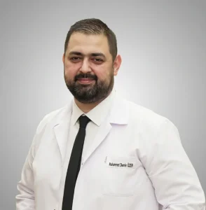
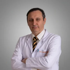
Optic neuropathy means damage to the optic nerve, the cable that carries everything you see from your eye to your brain. At Laser Istanbul Clinic, your optic nerve is assessed by a named eye surgeon: Doç. Dr. Muhammet Derda Özer (Associate Professor of Ophthalmology), Prof. Dr. Ahmet Satıcı or Op. Dr. Aysan Bayram, depending on the likely cause. You get a clear diagnosis, a written treatment plan and a personal interpreter from the first test to the last.

Optic neuropathy is any damage to the optic nerve. The main causes are inflammation (optic neuritis), poor blood supply (ischaemic optic neuropathy), glaucoma, pressure from a mass, toxins or vitamin deficiency, and inherited conditions. Treatment depends entirely on the cause. In Istanbul, Doç. Dr. Özer, Prof. Dr. Satıcı or Op. Dr. Bayram can examine your optic nerve, review your scans and give a written plan. Sudden vision loss needs same-day care wherever you are.
Sudden loss of vision: get seen today
If you lose vision suddenly in one or both eyes, see an eye doctor or go to an emergency department today, wherever you are. Do not wait to travel.
Be especially quick if you are over 50 and also have a new headache, a tender scalp, pain in your jaw when chewing, or feel generally unwell. These can be signs of giant cell arteritis (temporal arteritis), an inflammation of the arteries. According to the American Academy of Ophthalmology's EyeWiki, the sight loss it causes can spread to the second eye within days, and urgent high-dose steroids usually prevent that. It is treated as an emergency.
New optic neuritis also needs a prompt local examination, so that serious look-alikes are ruled out and treatment is not delayed. Our free online review is for planning and second opinions, not for new emergencies.
What does the optic nerve do? Function, anatomy and pathway
The optic nerve carries visual information from the retina to the brain, and it also carries the signal for the pupil's reaction to light. Without it, a perfectly healthy eye cannot see.
Optic nerve anatomy in brief. According to StatPearls (US National Library of Medicine), about 1.2 million nerve fibres from the retina's ganglion cells gather at the optic disc, the round spot at the back of the eye your optician sees with a light. The fibres leave the eye through a sieve-like plate called the lamina cribrosa, where they gain a fatty insulating coat (myelin) made by the same cells that insulate nerves in the brain. This is why the optic nerve counts as part of the central nervous system, and why diseases of the brain's myelin, such as multiple sclerosis, can affect it.
The optic nerve pathway, step by step:
- Retina: light-sensitive cells turn light into signals, which pass to the retinal ganglion cells.
- Optic nerve: the ganglion cell fibres travel from the back of the eye, through a bony channel (the optic canal), into the skull.
- Optic chiasm: the two optic nerves meet. Fibres from the inner (nasal) half of each retina cross to the other side; fibres from the outer half stay on the same side.
- Optic tracts: the regrouped fibres continue to the lateral geniculate nucleus, a relay station in the thalamus.
- Optic radiations: from there, signals fan out through the brain.
- Visual cortex: vision is finally processed in the occipital lobe at the back of the brain.
Because of this crossing, damage in front of the chiasm usually affects one eye, while damage at or behind it affects the same side of the visual field in both eyes. That pattern is exactly what a visual field test looks for.
What is optic neuropathy? The main types
Optic neuropathy is the medical term for damage to the optic nerve, whatever the cause. The hallmarks are blurred or dim vision, colours that look washed out, gaps in the visual field, an abnormal pupil reaction in the affected eye and, over time, a change in how the optic disc looks. The type tells your doctor which treatment, if any, can help.
| Type | What happens | Typical patient | Onset |
|---|---|---|---|
| Optic neuritis (inflammatory) | The immune system inflames the optic nerve | Adults aged 20–45, more often women | Over hours to days, often with pain on eye movement |
| Non-arteritic ischaemic optic neuropathy (NAION) | Poor blood flow to the front of the optic nerve | Usually over 50, with blood-pressure, diabetes or cholesterol problems | Sudden and painless, often noticed on waking |
| Arteritic ischaemic optic neuropathy | Giant cell arteritis blocks the nerve's small arteries | Over 50 | Sudden; an emergency |
| Glaucoma | Gradual loss of optic-nerve fibres, often with raised eye pressure | Risk rises with age | Slow and silent at first |
| Compressive | A tumour, enlarged eye muscles (thyroid eye disease) or another mass presses on the nerve | Any age | Usually slow and progressive |
| Toxic or nutritional | Medicines (for example the tuberculosis drug ethambutol), methanol, or low vitamin B12 or folate | Any age | Gradual, usually both eyes |
| Hereditary | Inherited faults, such as Leber hereditary optic neuropathy (LHON) | Young adults, mostly men in LHON | Painless, one eye then the other |
| Traumatic | Injury to the head or eye socket | Any age | After an injury |
Optic neuritis: symptoms, causes, treatment and recovery
Optic neuritis is inflammation of the optic nerve, and it typically affects young adults. EyeWiki puts its incidence at 1–5 per 100,000 people a year, most often between 20 and 45, with women affected about 3 times for every 2 men.
Optic neuritis symptoms
The typical optic neuritis eye shows these symptoms:
- Blurred, dim or "foggy" vision in one eye, developing over hours to days
- Pain behind the eye, worse when you move it. EyeWiki reports pain on eye movement in 87% of people
- Washed-out colours, especially red
- Patches missing from your vision, often in the centre
- Vision that worsens when you are hot, for example in a shower or during exercise (Uhthoff's phenomenon)
- Sometimes flashes of light
In about two-thirds of cases the optic nerve looks normal at first, because the inflammation sits behind the eye. EyeWiki notes that the optic disc is swollen in up to 35%. The pupil in the affected eye usually reacts less strongly to light, a sign doctors call a relative afferent pupillary defect.
Optic neuritis causes
Optic neuritis happens when the immune system attacks the myelin coat of the optic nerve. NHS patient information lists these causes and associations:
- Multiple sclerosis (MS). Optic neuritis can be the first sign of MS.
- Neuromyelitis optica spectrum disorder (NMOSD), linked to aquaporin-4 (AQP4) antibodies
- MOG antibody disease (MOGAD)
- Sarcoidosis and lupus, and other autoimmune diseases
- Infections, such as measles, mumps, chickenpox, syphilis, tuberculosis and Lyme disease
- No clear trigger, in many people
How is it linked to MS? In the long-term follow-up of the Optic Neuritis Treatment Trial (ONTT), half of the patients developed MS within 15 years. The brain MRI at the start made a large difference: 25% of people with no brain lesions developed MS, compared with 72% of those with one or more lesions. This is why an MRI scan is a standard part of the work-up.
How is optic neuritis diagnosed?
Your eye doctor checks your vision, colour vision, pupils, visual field and optic nerve, and usually orders an MRI scan of the brain and orbits with contrast. EyeWiki reports that MRI shows optic-nerve enhancement in 94% of cases. Blood tests for AQP4 and MOG antibodies are added when the picture is "atypical", for example:
- both eyes affected at once
- very severe vision loss at the start
- vision that keeps getting worse
- marked optic disc swelling with bleeding
- vision that does not start to recover
Optic neuritis treatment
Treatment of optic neuritis depends on the cause; for typical (MS-type) optic neuritis, high-dose steroids speed up recovery but do not change the final result. The ONTT, which enrolled 457 patients between 1988 and 1991, compared three options:
| ONTT treatment group | Result |
|---|---|
| Intravenous methylprednisolone, 1,000 mg a day for 3 days, then 11 days of oral prednisone | Faster recovery of vision; same final vision at 6 months as placebo |
| Oral prednisone alone (standard dose) | No benefit over placebo, and more recurrences, so it is not used on its own |
| Placebo | Most patients still recovered well |
UK NHS patient information reflects this: steroids are usually given into a vein for three days, sometimes by mouth, and are especially recommended when vision loss is severe or both eyes are affected. Steroids have side effects, which your doctor discusses with you before you start.
Atypical optic neuritis is treated differently and more urgently. The American Academy of Ophthalmology's EyeNet notes that NMOSD-related optic neuritis has a poorer visual outlook and is treated with high-dose steroids plus plasma exchange (a procedure that filters antibodies out of the blood), followed by long-term treatment to prevent attacks. MOGAD-related optic neuritis usually responds well to steroids and generally has a good visual outcome. These conditions are managed jointly with a neurologist.
Optic neuritis recovery time: how long does it last?
Most people with typical optic neuritis start to see better within a few weeks, and most of the recovery happens within 1–3 months. Improvement can continue for up to six months or longer. In the ONTT, 94% of patients had recovered to 20/40 vision or better at 5 years, and only 3% were at 20/200 or worse.
Even after a good recovery, many people notice that colours, contrast or vision in dim light are not quite the same as in the other eye. OCT scans often show some thinning of the nerve fibre layer from 6–8 weeks after the attack, a permanent record of the episode.
Other optic neuropathies: what each one means for you
Ischaemic optic neuropathy (NAION and giant cell arteritis)
NAION is the most common sudden optic neuropathy in people over 50. EyeWiki describes painless sight loss in one eye, often noticed on waking, in people who usually have a small, "crowded" optic disc and risk factors such as high blood pressure, diabetes, high cholesterol, smoking or sleep apnoea. There is no proven treatment to restore vision; a large US trial (IONDT) found that optic nerve sheath decompression surgery was not effective and may be harmful. About half of patients end up with 20/30 vision or better, and 15–24% develop it in the other eye within 5 years, so controlling your vascular risk factors matters.
In June 2025 the European Medicines Agency concluded that NAION is a very rare side effect of semaglutide (Ozempic, Wegovy, Rybelsus), affecting up to 1 in 10,000 people taking it. If you notice sudden or rapidly worsening vision while on these medicines, contact your doctor without delay; do not stop a prescribed medicine on your own.
Arteritic ischaemic optic neuropathy, caused by giant cell arteritis, is the emergency described at the top of this page. Blood tests (ESR and CRP) and often a temporal artery biopsy confirm it, but steroids are started straight away.
Glaucoma: a long-term optic neuropathy
The US National Eye Institute describes glaucoma as a group of eye diseases that damage the optic nerve. It has no early symptoms, and as many as half of people with glaucoma do not know they have it. Lost vision does not come back, but early treatment with drops, laser or surgery can often stop further damage. At our practice, glaucoma care is led by Prof. Dr. Ahmet Satıcı.
Compressive, toxic, nutritional and hereditary optic neuropathy
- Compressive optic neuropathy happens when something presses on the nerve: a tumour of or near the optic nerve (such as a meningioma or a pituitary lesion), enlarged eye muscles in thyroid eye disease, or another mass in the eye socket. EyeWiki lists these among the causes of an abnormal pupil reaction. Imaging finds the cause, and treating it can protect the nerve.
- Toxic and nutritional optic neuropathy. The American Academy of Ophthalmology highlights the tuberculosis medicine ethambutol, other drugs such as linezolid, methanol poisoning, and deficiency of vitamin B12 or folate. Caught early and with the cause removed, many patients recover some, though not always all, of their vision, slowly over months.
- Leber hereditary optic neuropathy (LHON) is a mitochondrial (maternally inherited) condition that usually causes painless sight loss in one eye, then the other, in young adults. With the most common mutation (m.11778G>A), men are affected about six times more often than women. Since September 2015, idebenone (Raxone) has been authorised in the EU to treat visual impairment from LHON in adolescents and adults.

Who should come to Istanbul for an optic nerve assessment?
An optic nerve assessment in Istanbul suits people with a non-urgent question about their optic nerve who want a thorough work-up and a clear answer from a named surgeon. That includes people who:
- have been told their optic nerve "looks pale", "swollen" or "cupped" and want a clear diagnosis
- had optic neuritis and want a review of recovery, OCT and visual fields, or a second opinion on the diagnosis
- have slowly worsening vision or colour vision that has not been explained
- have glaucoma, or suspected glaucoma, and want an independent assessment and treatment plan
- need an optic-nerve check before eye surgery, such as cataract or lens replacement
- live in Türkiye or are visiting, and want to be seen without a long wait
It is not the right first step if you have sudden vision loss, new pain on eye movement with blurring, or symptoms of giant cell arteritis. Get seen locally first. Your local reports, scans and blood results can then be sent to us for a second opinion.
How we diagnose optic neuropathy: tests and technology
A diagnosis of optic neuropathy rests on a few key findings: reduced vision, poorer colour vision, a pupil that reacts weakly to light in the affected eye and a change in the optic disc. Your surgeon builds on that with targeted tests.
| Test | What it shows | Why it matters |
|---|---|---|
| Visual acuity and colour vision | How sharp your vision is; how well you see colours | Colour vision is often affected early in optic-nerve disease |
| Pupil examination | A weaker reaction in one eye (relative afferent pupillary defect) | Points to the optic nerve rather than the lens or cornea |
| Dilated examination of the optic disc | Swelling, paleness, cupping, bleeding | Helps separate inflammation, ischaemia, glaucoma and pressure |
| Visual field test (perimetry) | Which parts of your vision are missing | The pattern helps locate the problem along the pathway |
| OCT scan of the optic nerve and macula | Thickness of the nerve fibre and ganglion cell layers | Measures damage, tracks change over time and rules out retinal causes |
| Visual evoked potentials (VEP) | How fast signals travel from eye to brain | A delayed signal supports optic-nerve disease |
| Eye pressure and drainage-angle check | Glaucoma risk | Glaucoma slowly damages the optic nerve, often without symptoms |
| Blood tests, MRI and specialist referral | Inflammation markers, antibodies, vitamin levels; brain and orbit imaging | Identifies causes outside the eye |
Technology we use: {{OCT, visual field and VEP/electrophysiology equipment actually used}}. {{Whether MRI scans and blood tests (AQP4/MOG antibodies, ESR/CRP, vitamin B12) can be arranged in Istanbul during your visit, and how neurology input is arranged when needed}}.
Optic neuropathy treatment by cause
There is no single optic neuropathy treatment: the aim is to treat the cause quickly, protect the fibres that are still working and support your vision. Lost optic-nerve fibres cannot currently be replaced.
| Cause | Main treatment (from the sources below) |
|---|---|
| Typical optic neuritis | High-dose intravenous steroids for faster recovery, especially when vision loss is severe; MRI and neurology review for MS risk |
| NMOSD or MOGAD optic neuritis | Urgent high-dose steroids, plasma exchange for NMOSD, long-term immune treatment with a neurologist |
| Giant cell arteritis | Immediate high-dose steroids to protect the other eye |
| NAION | No proven treatment to restore vision; control blood pressure, diabetes, cholesterol and sleep apnoea |
| Glaucoma | Pressure-lowering drops, laser or surgery |
| Compressive | Treat the mass, inflammation or infection, often with other specialists |
| Toxic or nutritional | Stop the toxic medicine or exposure (with your prescribing doctor), replace vitamins |
| LHON | Idebenone where appropriate; genetic counselling |
What about stem cells for the optic nerve? Stem cell treatments for the optic nerve are still experimental and only available in clinical trials. We do not offer them, and we suggest caution with anyone who sells them as a proven cure.

Optic nerve assessment in Istanbul: price and what is included
An optic nerve assessment with Laser Istanbul Clinic costs {{£ assessment price}}. If you need treatment, your surgeon confirms the full price in writing before anything is done.
Your assessment includes:
- a full eye examination with your named surgeon
- the optic-nerve tests your surgeon selects, such as OCT, visual field and, where needed, VEP
- a written report and treatment plan, in English
- a personal interpreter and a dedicated coordinator during your visit
- remote follow-up after you return home, so your local doctor can work from the same plan
If treatment is needed in Istanbul, the price includes the treatment by your named surgeon, medication and follow-up checks in Istanbul. Any procedure takes place in a fully equipped, Ministry of Health–licensed operating theatre in Istanbul.
Not included: flights, hotel and airport transfers. Hotel and transfers are a separate charge, and we can arrange them if you ask. {{Whether MRI, blood tests and any neurology consultation are included or charged separately, with prices}}.
Sending your reports first is free. Upload your eye reports, OCT and visual field printouts, and any MRI reports. A surgeon replies within {{response time}} and tells you whether a visit is worthwhile.
Your optic nerve surgeons

Your optic nerve assessment, step by step
Free online review
Send your eye reports, OCT, visual fields, MRI reports and a short description of your symptoms. A surgeon tells you whether a visit is worthwhile and which tests to plan.
Plan your dates
Allow {{typical stay}} in Istanbul for an assessment; longer if treatment is planned. Your coordinator books your appointments.
Examination and tests (day 1)
Vision, colour vision, pupils, eye pressure, dilated examination, OCT and visual field. Dilating drops blur your vision for a few hours, so do not drive.
Further tests if needed (day 1–2)
VEP and any blood tests or scans your surgeon requests.
Results and written plan
Your surgeon explains the diagnosis through your interpreter and gives you a written report and plan.
Treatment and home
If treatment is needed, it starts in Istanbul or is handed over to your local doctors. Your coordinator stays in touch, and your surgeon reviews progress remotely at agreed intervals.
What to expect after diagnosis and treatment
-
Day of the tests
Blurred near vision and light sensitivity for a few hours after dilating drops
-
First days of steroid treatment (if given for optic neuritis)
Recovery may begin; your doctor monitors side effects
-
2–4 weeks
In typical optic neuritis, vision often starts to improve
-
1–3 months
Most recovery from typical optic neuritis happens in this period
-
6–8 weeks onwards
OCT may show some permanent thinning of the nerve fibre layer, even when vision recovers
-
6–12 months
Slower improvement can continue; repeat OCT and visual fields set a new baseline
-
Long term
Regular checks for glaucoma; MS or NMOSD follow-up with a neurologist where relevant
Get urgent help if your vision gets worse again, the other eye is affected, or you develop new headache, jaw pain or weakness.

Outlook and limits: what treatment can and cannot do
The outlook after optic neuropathy depends on the cause and on how quickly it is treated. Typical optic neuritis has a good outlook: most people recover useful vision. NAION and giant cell arteritis usually leave permanent loss, which is why protecting the second eye matters so much. In glaucoma and toxic optic neuropathy, early treatment often stops further damage.
No operation can repair or replace a damaged optic nerve. Treatment protects what is left. Low-vision aids and visual rehabilitation help many people use their remaining vision well. Your surgeon will give you a realistic outlook for your eyes, without promises.
Optic neuropathy and optic neuritis: frequently asked questions
Optic neuropathy is damage to the optic nerve, the nerve that carries visual signals from the eye to the brain. It can be caused by inflammation, poor blood supply, glaucoma, pressure, toxins, vitamin deficiency, injury or inherited conditions. Typical signs are reduced vision, washed-out colours and gaps in the visual field. The treatment depends on the cause.
The first symptoms are usually pain behind one eye that is worse when you move it, followed within hours to days by blurred or dim vision. Colours, especially red, may look faded. Some people notice a dark patch in the centre of their vision or that vision worsens when they are hot. See an eye doctor promptly if you notice these.
In the UK, optic neuritis is usually assessed by an eye specialist with vision, colour and pupil tests, blood tests and often an MRI scan. NHS patient information says steroids are given into a vein for three days, or sometimes by mouth, mainly when vision loss is severe or both eyes are affected. Many people recover without treatment, because steroids speed recovery but do not change the final result in typical cases.
In typical optic neuritis, vision usually starts to improve within a few weeks, and most recovery happens within 1–3 months. Improvement can continue for six months or more. In the Optic Neuritis Treatment Trial, 94% of patients had 20/40 vision or better at five years, though colour and contrast may stay slightly different.
It can be, but not always. In the Optic Neuritis Treatment Trial, 50% of patients developed MS within 15 years: 25% of those with a normal brain MRI and 72% of those with one or more brain lesions. That is why an MRI scan and, where needed, a neurologist's review are part of the work-up.
Inflamed optic nerves often recover much of their function, as in typical optic neuritis. However, nerve fibres that have died do not grow back, so vision lost from NAION, giant cell arteritis or advanced glaucoma is usually permanent. Treatment aims to stop further damage and protect the other eye.
Yes. Sudden vision loss needs same-day assessment by an eye doctor or emergency department. If you are over 50 and also have headache, scalp tenderness or jaw pain when chewing, giant cell arteritis is possible, and urgent steroids can protect your other eye.
In June 2025 the European Medicines Agency concluded that NAION, a type of ischaemic optic neuropathy, is a very rare side effect of semaglutide, affecting up to 1 in 10,000 users. If you notice sudden or worsening vision while taking it, contact your doctor without delay. Do not stop a prescribed medicine without medical advice.
No. Stem cell treatments for the optic nerve are still experimental and available only within clinical trials. We focus on proven diagnosis and treatment, and we are open with you about what is and is not established.
With Laser Istanbul Clinic, an optic nerve assessment costs {{£ assessment price}}, including your examination, the tests your surgeon selects, a written report, an interpreter and remote follow-up. Hotel and transfers are extra. Sending your reports for a first review is free.
Sources for the facts on this page
- EyeWiki (American Academy of Ophthalmology), Demyelinating Optic Neuritis: incidence 1–5/100,000; age 20–45; female:male 3:2; pain on eye movement 87%; disc swelling in up to 35%; Uhthoff phenomenon, photopsias; MRI enhancement 94%; OCT thinning from 6–8 weeks; ONTT regimen (1,000 mg IV methylprednisolone × 3 days, then 11-day oral taper); oral prednisone alone linked to more recurrences; recovery mostly within 1–3 months; 94% ≥20/40 at 5 years; MS at 15 years 50% overall, 25% (no lesions) vs 72% (≥1 lesion); atypical features. https://eyewiki.org/Demyelinating_Optic_Neuritis
- ClinicalTrials.gov, Optic Neuritis Treatment Trial (ONTT), NCT00000146 (457 patients, 15 centres, recruited 1988–1991; three arms), with summary on EyeWiki Clinical Trials in Neuro-Ophthalmology: faster recovery with IV steroids, no difference at 6 months; oral prednisone no better than placebo; 3% ≤20/200 at 5 years. https://clinicaltrials.gov/study/NCT00000146 · https://eyewiki.org/Clinical_Trials_in_Neuro-Ophthalmology
- American Academy of Ophthalmology, What Is Optic Neuritis?: definition, symptoms (dim vision, faded colours, pain on eye movement, worse when hot), causes incl. MS and MOGAD, steroids as most common treatment. https://www.aao.org/eye-health/diseases/what-is-optic-neuritis
- NHS patient information on optic neuritis (Hull University Teaching Hospitals NHS Trust leaflet): symptoms, associated conditions (MS, neuromyelitis optica, sarcoidosis, lupus, measles, mumps, chickenpox, syphilis, tuberculosis, Lyme disease), tests, methylprednisolone recommended for severe or two-eye cases, recovery within weeks or months, "may be the first sign of MS". https://www.hey.nhs.uk/patient-leaflet/optic-neuritis/
- NHS patient information on optic neuritis (Great Ormond Street Hospital NHS Foundation Trust): immune attack on myelin, can follow viral infection; steroids into a vein for three days or by mouth; MRI and blood tests; recovery continuing up to six months. https://www.gosh.nhs.uk/conditions-and-treatments/conditions-we-treat/optic-neuritis/
- American Academy of Ophthalmology EyeNet, Optic Neuritis: NMOSD and MOGAD Treatment Evolves: AQP4 vs MOG antibodies; NMOSD-ON poorer outcome, steroids plus plasma exchange, long-term immunotherapy; MOGAD-ON usually steroid-responsive with favourable outcome. https://www.aao.org/eyenet/article/nmosd-and-mogad-treatment-evolves
- StatPearls (NCBI Bookshelf, US National Library of Medicine), Neuroanatomy, Cranial Nerve 2 (Optic): about 1.2 million retinal ganglion cell axons; myelination after the lamina cribrosa by oligodendrocytes; afferent pathway for vision and pupillary reflexes; pathway retina → optic nerve → chiasm (nasal fibres cross) → optic tracts → lateral geniculate nucleus → optic radiations → occipital visual cortex. https://www.ncbi.nlm.nih.gov/books/NBK507907/
- EyeWiki (AAO), Non-Arteritic Anterior Ischemic Optic Neuropathy (NAION): painless loss, often on waking; small "disc at risk"; vascular risk factors; no effective therapy; IONDT found decompression surgery not effective and possibly harmful; about 50% reach 20/30 or better; 15–24% fellow-eye involvement within 5 years; ESR/CRP and biopsy to exclude GCA. https://eyewiki.org/Non-Arteritic_Anterior_Ischemic_Optic_Neuropathy_(NAION)
- European Medicines Agency, PRAC concludes eye condition NAION is a very rare side effect of semaglutide medicines Ozempic, Rybelsus and Wegovy (June 2025): up to 1 in 10,000; contact doctor promptly with sudden vision loss. https://www.ema.europa.eu/en/news/prac-concludes-eye-condition-naion-very-rare-side-effect-semaglutide-medicines-ozempic-rybelsus-wegovy
- EyeWiki (AAO), Arteritic Anterior Ischemic Optic Neuropathy (AAION) and Giant Cell Arteritis: sudden, irreversible loss; fellow eye can be involved within days; ophthalmic emergency; urgent high-dose steroids usually prevent further loss. https://eyewiki.org/Arteritic_Anterior_Ischemic_Optic_Neuropathy_(AAION) · https://eyewiki.org/Giant_Cell_Arteritis
- American Academy of Ophthalmology, What Is Ischemic Optic Neuropathy?: risk factors, age 50+, vision loss usually permanent, temporal arteritis treated with steroids, low-vision aids. https://www.aao.org/eye-health/diseases/what-is-ischemic-optic-neuropathy
- American Academy of Ophthalmology EyeNet, Acquired Optic Neuropathy: First, Undo Harm: ethambutol, linezolid and other drugs, methanol, B12/folate deficiency; early removal of the cause allows partial recovery over months. https://www.aao.org/eyenet/article/acquired-optic-neuropathy-first-undo-harm
- US National Eye Institute, Glaucoma: group of eye diseases that damage the optic nerve; no early symptoms; up to half unaware; drops, laser, surgery; early treatment can often stop damage. https://www.nei.nih.gov/eye-health-information/eye-conditions-and-diseases/glaucoma
- EyeWiki (AAO), Leber Hereditary Optic Neuropathy, and GeneReviews (NCBI), Leber Hereditary Optic Neuropathy: maternal mitochondrial inheritance, sequential painless loss in young adults; m.11778G>A most common; male:female about 6:1 for this mutation. https://eyewiki.org/Leber_Hereditary_Optic_Neuropathy · https://www.ncbi.nlm.nih.gov/books/NBK1174/
- Idebenone (Raxone) EU authorisation September 2015 for visual impairment in adolescents and adults with LHON: European Medicines Agency product information and Lyseng-Williamson KA, Idebenone: A Review in Leber's Hereditary Optic Neuropathy, Drugs 2016. https://www.ema.europa.eu/en/documents/product-information/raxone-epar-product-information_en.pdf · https://pubmed.ncbi.nlm.nih.gov/27071925/
- EyeWiki (AAO), Relative Afferent Pupillary Defect: RAPD reflects unilateral or asymmetric retinal or optic nerve disease; causes include optic neuritis (MS, NMOSD, MOG), ischaemic optic neuropathies, compressive lesions (optic nerve glioma/meningioma, sphenoid wing meningioma, pituitary lesions, thyroid-related orbitopathy, orbital tumours), asymmetric glaucoma, inflammation, infection, hereditary, trauma, radiation. https://eyewiki.org/Relative_Afferent_Pupillary_Defect
Get a clear answer about your optic nerve
Send us your eye reports, OCT scans, visual fields and MRI reports. Doç. Dr. Özer, Prof. Dr. Satıcı or Op. Dr. Bayram will review them and tell you whether a visit to Istanbul would help, with no obligation.
3 eye surgeons
Your case is reviewed by a named surgeon, not a salesperson. Reply within {{response time}}.
 Prof. Dr. Ahmet Satıcı
 Doç. Dr. Muhammet Derda Özer
 Op. Dr. Aysan Bayram