
- Home
- Knowledge centre
- Keratoconus & cornea guides
- Keratoconus LASIK: Can Patients Have Laser Surgery or an ICL?
Keratoconus LASIK: Can Patients Have Laser Surgery or an ICL?
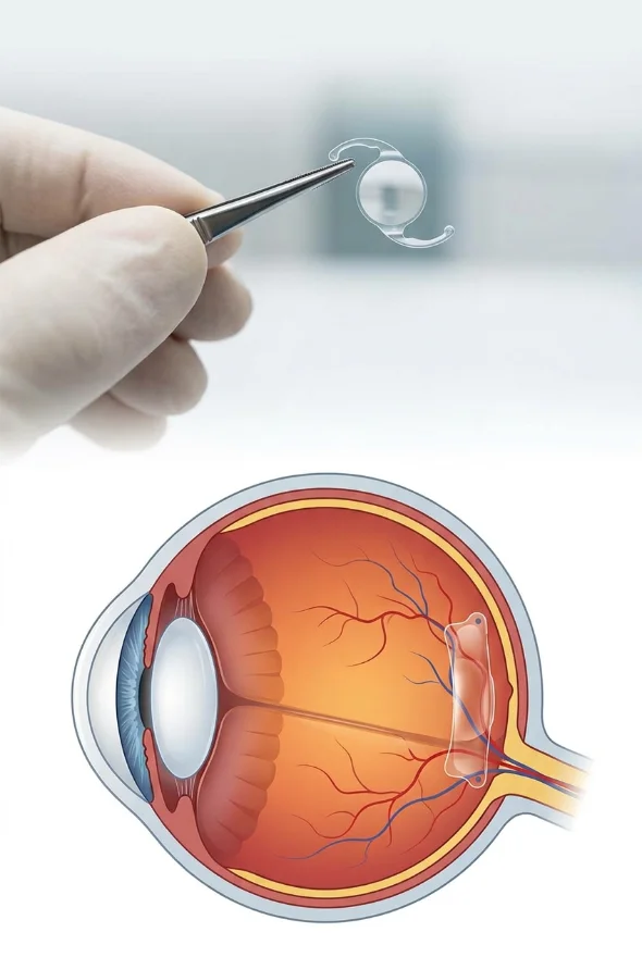
Keratoconus and LASIK do not mix: the FDA says laser vision correction has not been shown safe in keratoconus, and the American Academy of Ophthalmology's EyeWiki calls any refractive procedure a contraindication. An implantable collamer lens (ICL) can be considered once cross-linking has stabilised the cornea. At Laser Istanbul Clinic, cornea specialist Doç. Dr. Muhammet Derda Özer judges your cornea first, and Prof. Dr. Ahmet Satıcı advises on an ICL.
Can you have LASIK if you have keratoconus?
No. Standard LASIK is not recommended if you have keratoconus, because it makes a weak cornea weaker. Keratoconus is a condition in which the cornea, the clear dome at the front of the eye, gradually thins and bulges forward into a cone shape. EyeWiki, the AAO's clinical encyclopaedia, defines it as a progressive thinning and steepening of the cornea that causes irregular astigmatism and reduced visual quality, and it is usually found in both eyes, though often more in one.
The two main sources agree on LASIK:
- The US Food and Drug Administration (FDA) says the safety and effectiveness of refractive procedures has not been determined in people with some diseases, and names keratoconus among them. It also warns that performing a refractive procedure on a cornea that is too thin may cause blinding complications.
- EyeWiki states that any type of refractive procedure is considered a contraindication in keratoconic patients because of the unpredictability of the outcome.
That is why a careful clinic will not offer you LASIK on a keratoconic cornea, however much you want to be free of glasses or contact lenses. If you were told elsewhere that you are "not suitable for laser", the reason is very often exactly this.
Why is LASIK risky when you have keratoconus?
LASIK weakens the cornea in two ways. The surgeon cuts a thin flap across the front of the cornea, and the laser then removes tissue from underneath it to change the cornea's shape. In a healthy, thick cornea there is enough strong tissue left behind to hold the shape. In keratoconus the cornea is already thin and structurally weak, so removing more tissue can push it into progressive bulging.
That complication has a name: ectasia (also called keratectasia), a progressive thinning and steepening of the cornea after refractive surgery, the same shape change that keratoconus causes. Doctors have known since at least 1998 that it can happen when LASIK is done on an eye with an undetected early form of keratoconus: a case report in the Journal of Cataract & Refractive Surgery described ectasia after LASIK in a case of "forme fruste" (very early, hard-to-spot) keratoconus.
A 2021 systematic review of the world literature (Moshirfar et al., Ophthalmology and Therapeutics) gives a sense of the scale:
| Procedure | Reported ectasia in eyes without identifiable risk factors | Share of reported ectasia cases that had identifiable risk factors before surgery |
|---|---|---|
| LASIK | 90 per 100,000 eyes | 13.5% (228 of 1,681 eyes) |
| PRK (surface laser) | 20 per 100,000 eyes | 19% (13 of 70 eyes) |
| SMILE | 11 per 100,000 eyes | 42% (8 of 19 eyes) |
Source: Moshirfar et al. 2021. These rates are calculated from published case reports, so they are estimates, and they describe eyes that were not known to have keratoconus. They are not a guide to the risk in a keratoconic eye, which is much higher and not reliably predictable.
Two things stand out. First, LASIK had about 4.5 times the reported ectasia rate of PRK. Second, the authors point out that keratoconus is a spectrum, so pre-existing, unrecognised keratoconus may account for more post-surgery ectasia than the literature has assumed. The risk factors the review lists include a "keratoconus suspect" or forme fruste scan, pellucid marginal degeneration, a thin cornea and a thin layer of cornea left behind after the laser.
What this means for you: the safest approach is not to try to beat the odds with a different laser, but to confirm what your cornea is actually doing and treat that first.
What about SMILE and PRK for keratoconus?
Neither SMILE nor PRK on its own is a recommended treatment for keratoconus. They carry a lower reported ectasia risk than LASIK in healthy eyes (see the table above), but they still remove corneal tissue, and EyeWiki's contraindication covers "any type of refractive procedure" in keratoconus.
- SMILE has no flap, but it removes a disc of tissue from inside the cornea. The reported ectasia rate is the lowest of the three in healthy eyes, yet 8 of the 19 reported SMILE ectasia cases already had a recognised risk factor beforehand.
- PRK removes the surface layer and reshapes the front of the cornea. On its own it is also not a keratoconus treatment.
Is laser ever used together with cross-linking?
Sometimes, in carefully selected eyes. A specialist can combine a light, topography-guided surface laser (PRK) with corneal cross-linking (CXL), a procedure often called the Athens protocol. The aim is to smooth the irregular surface of the cone while the cross-linking strengthens the cornea, not to remove the prescription completely. The evidence comes from case series:
- In a 2009 series of 325 keratoconic eyes from one centre (Kanellopoulos, Journal of Refractive Surgery), 198 eyes had PRK and CXL on the same day and 127 had CXL first and PRK six months later. After an average of 36 months, uncorrected vision had improved from 0.96 to 0.30 logMAR with same-day treatment, against 0.90 to 0.49 with the staged approach, and the author concluded that same-day treatment appeared superior.
- A 2011 series of 31 eyes (Kymionis et al., American Journal of Ophthalmology) called the same-day approach "a promising treatment alternative" after 12 to 25 months.
These are consecutive series from specialist centres, not randomised trials, and the patients had progressive keratoconus; they were not people who simply wanted to stop wearing glasses. This combination is not a "keratoconus LASIK". It is a cornea-first procedure, and only your scans can say whether your cornea is a candidate.
Can keratoconus patients have laser or an ICL?
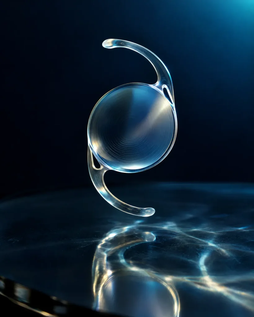
Laser, no; an ICL, yes in selected eyes. An implantable collamer lens (ICL) can be considered for a keratoconic eye that is stable, and it is generally done after corneal cross-linking. An ICL is a thin, soft lens placed inside the eye, behind the iris and in front of your natural lens. Nothing is cut from the cornea, so the cornea is not weakened further. That is the reason surgeons consider it when laser is ruled out.
An ICL corrects the focusing error (short sight and astigmatism) but it does not treat keratoconus itself. The cone is still there, and it is cross-linking that aims to keep it from getting worse.
What does the research on ICL in keratoconus show?
The evidence is encouraging but mostly observational (patients followed without a comparison group), with short to medium follow-up:
- A 2025 systematic review and meta-analysis in the American Journal of Ophthalmology pooled 16 observational studies covering 397 keratoconic eyes. Uncorrected vision, spherical equivalent, cylinder and refractive astigmatism improved significantly, no significant adverse events occurred, and the authors concluded ICLs were safe and effective in keratoconus. They also noted that the improvement in corrected vision (the vision you reach with glasses) was not statistically significant, and that the studies were observational with limited follow-up.
- A 2025 meta-analysis of ICL after cross-linking (7 observational studies, International Ophthalmology) found a mean gain in uncorrected vision of 1.27 logMAR at 6 months, no significant change in corrected vision, and no adverse effect on corneal curvature after cross-linking.
- A 2023 meta-analysis of 23 studies and 464 eyes (International Ophthalmology) found that implantable (phakic) lenses were comparably safe and effective, and that results improved when they were combined with cross-linking or corneal ring segments.
- A 2015 study of 30 eyes (Antonios et al., Journal of Ophthalmology) followed patients who had cross-linking and, six months later, a toric ICL (a version that also corrects astigmatism). At 12 months the average spherical equivalent fell from −8.37 to −1.36 dioptres, uncorrected vision improved from 1.57 to 0.17 logMAR, and 60% of eyes saw 20/30 or better without glasses. There were no cases of lens rotation, cataract, haze or infectious keratitis; a temporary rise in eye pressure in the first week was controlled with drops. In the 10 eyes followed to 24 months, vision stayed stable. It was a small retrospective study.
- A 2024 network meta-analysis (Niazi et al., Survey of Ophthalmology) suggested that, for keratoconic corneas with good corrected vision and a disease stage no higher than 3, an implantable lens combined with corneal ring segments and cross-linking could be the best approach, while irregular corneas did better with surface ablation, rings and cross-linking at longer follow-up.
Who is a suitable candidate for an ICL with keratoconus?
There is no single universal checklist. As an example of how strictly suitable eyes are chosen, the 2015 study above required:
| Check | Requirement in that study |
|---|---|
| Keratoconus stability | Refraction stable within ±0.50 dioptres after cross-linking |
| Cornea | Central thickness of at least 450 µm and average curvature (K reading) no higher than 56 dioptres |
| Space inside the eye | Anterior chamber depth of at least 2.8 mm |
| Endothelium (the cornea's inner cell layer) | More than 2,200 cells per mm² |
| Vision | Best-corrected vision better than 20/40, and intolerance of hard contact lenses |
Your own surgeon will use their own criteria, but these show why detailed scans matter. In short, the cornea must have stopped changing, there must be enough room inside the eye, and the inner cell layer must be healthy.
Why does cross-linking come first?
Because cross-linking is the treatment that aims to stop keratoconus from progressing, and any vision correction is safer and more predictable on a cornea that has stopped changing. Corneal cross-linking (CXL) uses riboflavin (vitamin B2) drops and ultraviolet light to strengthen the cornea's collagen. EyeWiki describes it as a minimally invasive procedure that prevents progression of corneal ectasia rather than correcting vision directly, and lists the US FDA approval date as 18 April 2016.
In an Australian controlled study reported by EyeWiki, after 36 months the cornea of treated eyes had flattened by an average of 1.03 dioptres, while untreated eyes had steepened by 1.75 dioptres, and 39% of the untreated group worsened by 2 dioptres or more.
How do surgeons judge whether the cornea is stable or progressing? A 2024 review in Community Eye Health (Deshmukh and Gore) says an increase in keratometry (curvature) of more than one dioptre, or a loss of corneal thickness of more than 15 microns, might be considered significant, provided the change is larger than the repeatability of the scanner. The same review notes that in younger patients, clinicians may offer cross-linking at diagnosis, without waiting for documented progression. Keratoconus usually starts in the late teens to early twenties (AAO) and can keep progressing into the mid-20s and 30s (EyeWiki), which is why surgeons wait until the cornea has settled before adding an ICL.
For the full picture of the treatment, see our page on Keratoconus treatment in Turkey. For teenagers, see Keratoconus in children and teenagers: why early CXL matters.
Can I have laser or an ICL if my scan only says "suspect" keratoconus?
Possibly, but only after a specialist has studied your corneal scans. "Keratoconus suspect" and "forme fruste keratoconus" are terms for corneas that show early or borderline warning signs without full keratoconus. The Moshirfar review names them, along with scan patterns such as an asymmetric bow-tie, inferior steepening and a skewed radial axis, as risk factors for ectasia after refractive surgery.
Because these corneas sit between normal and keratoconic, they are exactly where a misjudged laser treatment can do harm. Many surgeons prefer a non-corneal option such as an ICL, or a period of monitoring, in an eye with warning signs. The decision should rest on corneal topography and tomography (3D scans of the front and back surface of the cornea), thickness maps and a history of whether your prescription has been changing.
Options at a glance: laser, ICL and the alternatives
| Option | Is it used in keratoconus? | What it does | Key point |
|---|---|---|---|
| LASIK | No: not recommended | Reshapes the cornea under a flap | Weakens a thin cornea; risk of ectasia |
| SMILE | No, on its own | Removes a disc of tissue through a small cut | No flap, but still removes corneal tissue |
| PRK on its own | No | Reshapes the surface | Not a keratoconus treatment |
| Topography-guided PRK plus CXL | Selected eyes only, by a specialist | Smooths the cone while strengthening the cornea | Evidence from case series |
| ICL (often a toric ICL) | Selected stable eyes, usually after CXL | Lens inside the eye corrects the prescription | Cornea untouched; does not treat the cone |
| Corneal cross-linking (CXL) | Yes: the first step if progressing | Strengthens the cornea | Aims to stop progression, not to correct vision |
| Corneal ring segments | Mild to moderate keratoconus, contact-lens intolerance | Reshapes the cone | Needs adequate corneal thickness |
| Specialist contact lenses | Yes | Rigid lenses create a smooth optical surface | Often the first vision correction |
| Corneal transplant | Advanced keratoconus or scarring | Replaces part or all of the cornea | See our guide on corneal transplant success rates and recovery |
EyeWiki says corneal ring segments are approved for mild to moderate keratoconus in contact-lens-intolerant patients, with a corneal thickness above 450 µm at the insertion site. Deshmukh and Gore note that cross-linking has led to a reduction in the need for corneal transplant surgery.

What should you expect after an ICL in a keratoconic eye?
Expect better vision without glasses, but not necessarily perfect vision. In the studies above, the biggest gains were in uncorrected vision, while the vision reached with the best glasses or lenses changed little or not significantly. Because the cone is still present, the cornea's irregular shape can still limit how sharp your vision gets, and some people continue to wear glasses or specialist contact lenses for the best result.
Risks of any ICL include a temporary rise in eye pressure, as seen in the 2015 study, and the other complications of intraocular surgery. Your surgeon will go through these with you, and will check you again if your keratoconus changes. Keratoconus usually affects both eyes, so the cornea needs continued monitoring.
How do you find out which option suits you?
You need a specialist assessment of your corneal scans. The surgeon will want:
- Your recent corneal topography or tomography (the scan images, not only the report), and older ones if you have them, to show whether the cone is changing.
- Corneal thickness maps, including the thinnest point.
- Your prescription history, to see whether it has been stable.
- Details of any previous cross-linking, rings or contact lens use.
- Measurements of the space inside the eye and of the cornea's inner cell layer, if an ICL is being considered.
You can send these for a free review before you travel: Free online consultation (upload prescription & scans). At Laser Istanbul Clinic, Doç. Dr. Muhammet Derda Özer, who treats keratoconus, cross-linking and corneal transplants, looks at your cornea and its stability. Prof. Dr. Ahmet Satıcı, who performs ICL and laser surgery, advises on whether an ICL or laser makes sense once your cornea is stable. If neither is right for you, you are told so plainly. Any surgery takes place in a fully equipped, Ministry of Health–licensed operating theatre in Istanbul.
Frequently asked questions
No. LASIK is not recommended in keratoconus, because it thins and weakens a cornea that is already thin and can lead to ectasia, a progressive bulging of the cornea. The FDA says the safety and effectiveness of refractive procedures have not been determined in people with keratoconus, and EyeWiki calls any refractive procedure a contraindication.
Standard laser eye surgery (LASIK, SMILE or PRK on its own) is not recommended. In selected eyes, a specialist may combine a gentle, topography-guided PRK with cross-linking, but that is a cornea-strengthening treatment first and a vision-correction treatment second. Your scans decide.
Yes, in selected eyes that are stable and have enough space and healthy inner cells. It is usually done after cross-linking has stabilised the cornea. A 2025 meta-analysis of 16 observational studies (397 eyes) found ICLs in keratoconus safe and effective, with the limits that the studies were observational and the follow-up short.
For a keratoconic eye, an ICL is generally the safer of the two, because it does not remove corneal tissue. But it does not treat the keratoconus, and it will not fix the irregular shape of the cornea. Cross-linking is what addresses the disease.
SMILE on its own is not recommended in keratoconus. It has no flap, but it still removes corneal tissue, and 8 of the 19 reported cases of ectasia after SMILE in a 2021 review already had a recognised risk factor before surgery.
Usually yes, if your keratoconus is progressing. Cross-linking aims to stop progression, and surgeons generally want the cornea stable before they implant an ICL. In the 2015 study, the ICL was implanted six months after cross-linking and only in eyes whose refraction had stayed within ±0.50 dioptres.
It carries a risk. Forme fruste keratoconus is an early, hard-to-spot form, and suspect scan patterns are recognised ectasia risk factors. Many surgeons prefer an alternative to laser in these eyes. A specialist should review your tomography before any decision.
You may. An ICL mainly reduces your short sight and astigmatism, but the irregular cornea can still limit the sharpness of your vision, so some people still use glasses or specialist contact lenses for their best sight.
First stop progression (cross-linking if the cornea is changing), then use specialist contact lenses, rings, an ICL or, in selected cases, a combined surface laser with cross-linking. Which one is best depends on your cornea's stage and thickness, which only your scans show.
Keratoconus, and wondering whether laser or an ICL is possible?
Send us your scans. Doç. Dr. Muhammet Derda Özer reviews your cornea and its stability, and Prof. Dr. Ahmet Satıcı advises on an ICL or laser once it is stable. You get a plain answer on which steps make sense for your eyes, and which do not.
Sources for the facts on this page
- All opened and checked on 7 Oct 2026 (publisher pages, PMC articles or Europe PMC abstracts).
- US Food and Drug Administration. When is LASIK not for me? https://www.fda.gov/medical-devices/lasik/when-lasik-not-me
- EyeWiki (American Academy of Ophthalmology). Keratoconus. https://eyewiki.org/Keratoconus
- EyeWiki (American Academy of Ophthalmology). Corneal Collagen Cross-Linking. https://eyewiki.org/Corneal_Collagen_Cross-Linking
- American Academy of Ophthalmology. What Is Keratoconus? https://www.aao.org/eye-health/diseases/what-is-keratoconus
- Moshirfar M, Tukan AN, Bundogji N, et al. Ectasia After Corneal Refractive Surgery: A Systematic Review. Ophthalmology and Therapeutics 2021 (PMID 34417707). https://pmc.ncbi.nlm.nih.gov/articles/PMC8589911/
- Seiler T, Quurke AW. Iatrogenic keratectasia after LASIK in a case of forme fruste keratoconus. Journal of Cataract & Refractive Surgery 1998 (PMID 9682124). https://pubmed.ncbi.nlm.nih.gov/9682124/
- Alkhabbaz AA, Karam MH, Pollmann AS, et al. Safety and Efficacy of Posterior Chamber Phakic Implantable Collamer Lenses in Patients with Keratoconus: A Systematic Review and Meta-Analysis. American Journal of Ophthalmology 2025 (PMID 39613132). https://pubmed.ncbi.nlm.nih.gov/39613132/
- Elgazzar AF, Tharwat E, Diab S, et al. Efficacy of implantable Collamer lens after corneal cross-linking in patients with keratoconus: a systematic review and meta-analysis. International Ophthalmology 2025 (PMID 40455349). https://pubmed.ncbi.nlm.nih.gov/40455349/
- Fard AM, Patel SP, Nader ND. The efficacy of 2 different phakic intraocular lens implant in keratoconus as an isolated procedure or combined with collagen crosslinking and intra-stromal corneal ring segments: a systematic review and meta-analysis. International Ophthalmology 2023 (PMID 37470861). https://pubmed.ncbi.nlm.nih.gov/37470861/
- Antonios R, et al. Safety and Visual Outcome of Visian Toric ICL Implantation after Corneal Collagen Cross-Linking in Keratoconus: Up to 2 Years of Follow-Up. Journal of Ophthalmology 2015. https://pmc.ncbi.nlm.nih.gov/articles/PMC4383407/
- Niazi S, Doroodgar F, Hashemi Nazari S, et al. Refractive surgical approaches to keratoconus: A systematic review and network meta-analysis. Survey of Ophthalmology 2024 (PMID 38710236). https://pubmed.ncbi.nlm.nih.gov/38710236/
- Kanellopoulos AJ. Comparison of sequential vs same-day simultaneous collagen cross-linking and topography-guided PRK for treatment of keratoconus. Journal of Refractive Surgery 2009 (PMID 19772257). https://pubmed.ncbi.nlm.nih.gov/19772257/
- Kymionis GD, Portaliou DM, Kounis GA, et al. Simultaneous topography-guided photorefractive keratectomy followed by corneal collagen cross-linking for keratoconus. American Journal of Ophthalmology 2011 (PMID 21794846). https://pubmed.ncbi.nlm.nih.gov/21794846/
- Deshmukh R, Gore D. Keratoconus: diagnosis and management. Community Eye Health 2024. https://pmc.ncbi.nlm.nih.gov/articles/PMC11921258/