
- Home
- Knowledge centre
- Eye glossary A–Z
- Retinal Detachment: Symptoms, Causes, Diagnosis and Treatment
Retinal Detachment: Symptoms, Causes, Diagnosis and Treatment
Medically reviewed by {{surgeon}}, {{surgeon's title and specialty}}, on {{review date}}. Last updated {{date}}.
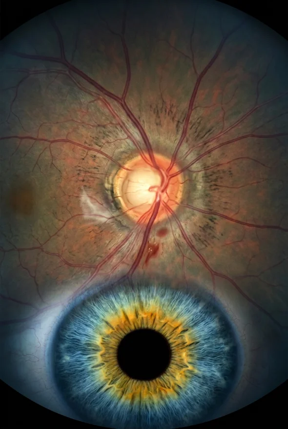
Retinal detachment (noun): a retinal detachment, or detached retina, happens when the retina, the thin light-sensitive layer at the back of the eye, lifts away from the wall of the eye. A detached retina cannot work properly, so vision blurs or is lost where it has come away. It is a medical emergency: it needs to be seen and treated quickly to protect sight, and it is treated with surgery. At Laser Istanbul Clinic, retinal conditions are managed by Doç. Dr. Muhammet Derda Özer (Doç. means Associate Professor) and Op. Dr. Aysan Bayram.
Retinal detachment: key facts
- What it is
- The retina pulls away from the back wall of the eye, so it can no longer send clear images to the brain
- Warning signs
- Sudden new floaters, flashes of light, a dark shadow or curtain across part of your vision
- Is it an emergency?
- Yes. The NHS tells people to call 111 straight away; the American Academy of Ophthalmology says to call an ophthalmologist right away
- Most common cause
- Age-related changes in the gel inside the eye (posterior vitreous detachment), and eye injury
- Higher risk if
- You are short-sighted, have had eye surgery such as cataract surgery, have had an eye injury, or have a family history
- Treatment
- Surgery: laser or freezing for tears, or pneumatic retinopexy, scleral buckle or vitrectomy for a detachment
- Outlook
- Treatment is successful for about 9 out of 10 people (National Eye Institute); some need a second operation
- Can it be prevented?
- No, according to the NHS, but spotting symptoms early allows prompt treatment
- Specialist
- Retina specialist (ophthalmologist); at Laser Istanbul Clinic, Doç. Dr. Muhammet Derda Özer and Op. Dr. Aysan Bayram
What is retinal detachment?
The retina lines the inside back wall of the eye and works like the sensor of a camera: it turns light into signals that the optic nerve carries to the brain. In retinal detachment, the retina is pulled away from its normal position. The National Eye Institute describes it as the retina being "pulled away from its normal position", and the American Academy of Ophthalmology explains that a detached retina does not work, which makes vision blurry.
The usual chain of events starts inside the eye. The eye is filled with a clear gel called the vitreous. With age the vitreous shrinks and may stick to the retina. When the pulling becomes strong enough, it can tear the retina, and fluid then passes through the tear and lifts the retina off the eye wall. A retinal detachment is therefore often the end point of a tear that was not found or treated in time, which is why tears and detachments are treated as one urgent problem.
What are the types of retinal detachment?
There are three types, each caused by a different mechanism.
| Type | What happens | Typical causes and risk factors |
|---|---|---|
| Rhegmatogenous (from the Greek for "break") | A tear or hole lets fluid pass under the retina and lift it | Posterior vitreous detachment, lattice degeneration (thin areas in the peripheral retina), high short-sightedness, previous eye surgery, injury |
| Tractional | Scar tissue on the retina's surface pulls the retina away | Proliferative diabetic retinopathy is the most common cause; also retinal vein occlusion and sickle cell retinopathy |
| Exudative | Fluid collects under the retina without a tear | Inflammation inside the eye (such as uveitis), tumours in the eye, abnormal blood vessels, and some general illnesses such as severe high blood pressure |
Most of this page describes the rhegmatogenous type, the one people usually mean by "a detached retina". Tractional detachments relate closely to diabetes; see Glossary: Diabetic retinopathy. Exudative detachments are managed by treating their cause, for example the inflammation or the tumour.
Is a retinal tear the same as a retinal detachment?
No. A retinal tear is a break in the retina. A detachment is when fluid collects through that break and the retina separates from the wall of the eye. A tear can often be sealed with a quick laser or freezing treatment before it becomes a detachment, which is one of the main reasons to be seen urgently when symptoms start.
Retinal detachment symptoms: what does a detached retina look like?
The warning signs usually appear suddenly. The National Eye Institute, the NHS and the American Academy of Ophthalmology list the same group:
- A lot of new floaters: small dark spots, lines, specks or cobwebs that drift across your vision
- Flashes of light in one eye or both, which can appear suddenly
- A dark shadow or curtain moving in from the side, or in the middle, of your field of vision
- Sudden blurred vision or other sudden changes in sight
Not every floater means trouble, and most floaters and flashes are harmless. The difference is newness and number: a sudden shower of new floaters, new flashes or a spreading shadow needs checking the same day. The National Eye Institute also notes that a small detachment may cause no symptoms at first, which is a reason to take any new change seriously and to have dilated eye checks if you are at higher risk.
What the symptoms can mean
| What you notice | What it can point to |
|---|---|
| Sudden shower of new floaters | The vitreous gel pulling away from the retina (posterior vitreous detachment) or a retinal tear |
| Flashes of light | The gel tugging on the retina |
| A shadow or grey curtain that grows | A retinal detachment that is spreading |
| Central vision suddenly blurred or distorted | The detachment may have reached the macula, the central part of the retina |

What causes retinal detachment and who is at risk?
The NHS says a detached retina is usually caused by changes to the jelly inside the eye as you get older (posterior vitreous detachment, or PVD), and adds that it is not clear exactly why PVD leads to retinal detachment in some people. The National Eye Institute names ageing and eye injury as the most common causes.
You are more likely to develop a detached retina if you:
- are short-sighted (myopic); the more severe the short-sightedness, the higher the risk, and EyeWiki lists myopia stronger than about -6 dioptres as a risk factor
- have had eye surgery, including cataract surgery (the NHS, the National Eye Institute and the American Academy of Ophthalmology all list this), or glaucoma surgery
- have had a serious eye injury
- have had a retinal detachment before, in either eye, or have a family history of it
- have a weak area in the retina found on examination, such as lattice degeneration or retinoschisis
- have diabetic retinopathy (mainly the tractional type)
EyeWiki gives the overall frequency as roughly 1 in 10,000 people, higher in men, in short-sighted people and after cataract surgery.
Can retinal detachment be prevented? According to the NHS there is nothing you can do to prevent it, but recognising the symptoms early means treatment can start quickly. If you are in a higher-risk group, regular dilated eye examinations let a specialist find weak areas or tears early, and the NHS notes that a detachment can happen more than once.
How is retinal detachment diagnosed?
Diagnosis is usually quick and done in the clinic. According to the American Academy of Ophthalmology, the National Eye Institute and EyeWiki:
Dilated eye examination
Drops widen the pupil, and the ophthalmologist uses specialised magnifying equipment to look at the whole retina, including its edges. EyeWiki describes indirect ophthalmoscopy as the gold standard for finding and locating retinal breaks.
Ultrasound (B-scan)
If bleeding or a cloudy lens stops the doctor seeing the retina, ultrasound shows whether it is detached.
Optical coherence tomography (OCT)
An OCT scan gives a detailed cross-section of the retina, confirms fluid under it and helps to tell a detachment from other retinal problems.
The surgeon also checks which part of the retina is involved. EyeWiki points out that whether the macula (the centre of the retina, which gives sharp reading and face vision) is still attached before surgery strongly affects the final vision, and that early detection and prompt repair are the strongest factors for a good result.
Retinal detachment treatment: how is a detached retina repaired?
A detached retina is treated with surgery. According to the NHS and the National Eye Institute, the choice depends on the type and size of the problem. EyeWiki adds that it also depends on whether the macula is involved and on the state of the lens.
| Treatment | What it does | When it is typically used |
|---|---|---|
| Laser or freezing (cryotherapy) treatment | Seals a retinal tear so that fluid cannot get behind the retina | Small tears, before a detachment has formed |
| Pneumatic retinopexy | A gas bubble is injected into the eye to push the retina back against the wall; you must hold a strict head position while it heals | A single or limited break in the upper part of the retina, a clear view into the eye, and a patient who can keep the required position (EyeWiki) |
| Scleral buckle | A small silicone or plastic band is attached to the outside of the eye to push the wall of the eye against the retina; it is usually permanent | Often chosen for younger patients who still have their natural lens, with peripheral breaks and no significant scar tissue (EyeWiki) |
| Vitrectomy | The vitreous gel is removed from the eye and replaced with gas, air or silicone oil that holds the retina in place while it heals | Patients with an artificial lens (after cataract surgery), a cloudy view, breaks near the back of the retina, scarring (PVR) or giant tears (EyeWiki) |
Some detachments are repaired with a combination of methods. Most operations are done under local anaesthetic, so you are awake while the eye is numbed, and the NHS says you do not usually need to stay in hospital overnight.
What goes into the eye after surgery?
After a vitrectomy the eye is filled with a gas bubble, air or silicone oil. A gas or air bubble slowly absorbs on its own. Silicone oil does not, and needs a second operation later to take it out. The bubble is why surgeons ask patients to hold a certain head position, up to 7 days according to the NHS, so that it presses on the right area of the retina.
What are the risks and success rate?
The National Eye Institute says treatment is ultimately successful for about 9 out of 10 people, and EyeWiki reports anatomical success of over 90% with modern techniques. Success means the retina has been reattached; it does not mean vision returns to normal in every case. The American Academy of Ophthalmology states that about 1 in 20 patients needs repeat surgery and lists these risks of surgery:
- infection and bleeding inside the eye
- raised eye pressure or glaucoma
- cataract (clouding of the natural lens)
- re-detachment
- permanently reduced vision even though the retina is reattached
The most common reason for a repair to fail is proliferative vitreoretinopathy (PVR), scar tissue that forms on the retina; EyeWiki says it occurs in about 8–10% of patients. Your surgeon should discuss these risks with you before the operation, and no surgeon can guarantee the result.
Recovery after retinal detachment surgery
| Topic | What to expect |
|---|---|
| First weeks | The NHS says to expect 2–6 weeks of blurred vision, with the eye possibly sore and red; you may need time off work and may not be allowed to drive |
| Positioning | If a gas bubble was used, you may have to lie or sit in a particular position for up to 7 days (NHS) |
| Vision improvement | The American Academy of Ophthalmology says vision starts to improve around 4–6 weeks after surgery, but it may take months or even years to settle |
| Flying and altitude | Not with a gas or air bubble in the eye. The American Academy of Ophthalmology advises against flying and high-altitude travel while a gas or air bubble is present |
| Follow-up | Regular checks are needed. Contact the hospital or A&E if pain, redness or blurring gets worse after surgery (NHS) |
Because of the flying restriction, ask your surgeon how long a bubble will last before you plan any journey, and never travel by plane with a gas or air bubble in the eye.

When to see a doctor about a retinal detachment
Get urgent, same-day care from an eye doctor or emergency eye service if you notice:
- a sudden shower of new floaters, or a big increase in floaters
- new flashes of light
- a dark shadow, or a grey curtain, covering part of your vision
- sudden blurred vision or other sudden vision changes
- after eye surgery: pain, redness or blurring that is getting worse
In the UK the NHS advises calling NHS 111 immediately; in the US the American Academy of Ophthalmology advises calling an ophthalmologist right away. Do not wait to see whether it settles, and do not wait for a planned trip: if you have these symptoms now, go to the nearest eye emergency service first.
Book an eye examination soon if you:
- are very short-sighted, have had cataract or other eye surgery, or have a family history of retinal detachment, and have not had a dilated retinal check
- have already had a retinal detachment in one eye (it can happen again)
- have diabetes and have not had a recent retinal screening
If you have already been diagnosed, treated or told you need surgery and want a second opinion, you can send your scans and reports for review.
Retinal detachment treatment with Laser Istanbul Clinic
Retinal conditions, including retinal detachment and vitrectomy, are the field of two of our three doctors:
- Doç. Dr. Muhammet Derda Özer (retina, medical and surgical; uveitis; ocular oncology; cornea). He speaks Turkish and English.
- Op. Dr. Aysan Bayram (cataract, laser and retinal disease). She speaks English and is a female surgeon, which some patients prefer to choose.
Surgery takes place in a fully equipped, Ministry of Health–licensed operating theatre in Istanbul. If you have already had a scan or a diagnosis, the doctors can review your reports before you decide anything. Because an acute retinal detachment is a same-day emergency, it should never be delayed for travel: if your symptoms have just started, see a local eye emergency service first. Our review is most useful for second opinions, planned surgery and aftercare planning.
Frequently asked questions
Retinal detachment is when the retina, the light-sensitive layer at the back of the eye, pulls away from the wall of the eye. The detached part stops working, so vision blurs or is lost there. It is an emergency that needs surgery.
The main warning signs are a sudden shower of new floaters, flashes of light, and a dark shadow or curtain moving across your vision. Sudden blurred vision can also occur. Any of these needs same-day assessment by an eye doctor.
Yes. Surgery reattaches the retina in about 9 out of 10 people, according to the National Eye Institute. About 1 in 20 patients needs a repeat operation, and some vision may be permanently reduced even after a successful repair, so early treatment matters.
Small tears can be sealed with laser or freezing treatment. A detachment is repaired with pneumatic retinopexy (gas bubble), a scleral buckle (band around the eye) or vitrectomy (removing the gel and replacing it with gas, air or silicone oil). Your surgeon chooses the method according to the type and position of the problem.
Expect 2–6 weeks of blurred vision, a sore or red eye and time off work, according to the NHS. You may need to hold a head position for up to 7 days if a gas bubble was used. Vision starts to improve at about 4–6 weeks and may keep improving for months.
Not while there is a gas or air bubble in the eye, according to the American Academy of Ophthalmology. Your surgeon will tell you when the bubble has gone and when it is safe to fly. If silicone oil was used, ask your surgeon about travel.
According to the NHS, it cannot be prevented. What you can do is recognise the warning signs early and get help immediately. If you are short-sighted, have had eye surgery or have a family history, regular dilated eye checks let a specialist find weak areas or tears early.
Yes. A retinal tear can lead to a detachment, and the same warning signs apply. Tears can often be sealed with laser or freezing treatment, which is a smaller procedure than detachment surgery.
No. A detached retina is the retina lifting away from the back of the eye and is a sudden emergency. Macular degeneration is a different, gradual disease of the central retina; see Glossary: Macular degeneration.
Sources for the facts on this page
- NHS. Detached retina. https://www.nhs.uk/conditions/retinal-detachment/ (checked 7 Oct 2026)
- NHS. Detached retina: treatment. https://www.nhs.uk/conditions/retinal-detachment/treatment/ (checked 7 Oct 2026)
- National Eye Institute (NIH). Retinal Detachment. https://www.nei.nih.gov/learn-about-eye-health/eye-conditions-and-diseases/retinal-detachment (last reviewed 5 Nov 2025)
- American Academy of Ophthalmology. What Is a Detached or Torn Retina? https://www.aao.org/eye-health/diseases/detached-torn-retina
- American Academy of Ophthalmology. What Are Floaters and Flashes? https://www.aao.org/eye-health/diseases/what-are-floaters-flashes
- EyeWiki (American Academy of Ophthalmology). Retinal Detachment. https://eyewiki.org/Retinal_Detachment
Been told you have a retinal tear or detachment, or want a second opinion?
Send us your scans and eye report. Doç. Dr. Muhammet Derda Özer or Op. Dr. Aysan Bayram will review them and explain which treatments may suit your eyes, with no obligation. If you have sudden new floaters, flashes or a shadow in your vision right now, go to your nearest eye emergency service first.
3 eye surgeons
Your case is reviewed by a named surgeon, not a salesperson. Reply within {{response time}}.
 Prof. Dr. Ahmet Satıcı
 Doç. Dr. Muhammet Derda Özer
 Op. Dr. Aysan Bayram