

Sclera
The white, outer wall of the eye, which the cornea joins at the limbus.
Glossary: ScleraThe clear window at the front of the eye: what it is made of, what it does, which conditions affect it, how it is examined and how it is treated.

Cornea (noun, plural corneas): the clear, dome-shaped window at the front of the eye, covering the coloured iris and the pupil. It protects the inside of the eye and does most of the eye's focusing, bending incoming light so that it can be focused onto the retina. The cornea has no blood vessels, which keeps it transparent, and it is one of the most sensitive tissues in the body.
The front surface of the eye, in front of the iris and pupil; it joins the white of the eye (the sclera) at a ring called the limbus.
None. Nutrients and oxygen come from the tears on the outside and the fluid inside the eye (aqueous humour).
The cornea looks like a simple clear dome, but its structure is precise: each layer has its own job, and how well a layer heals decides what happens after an injury or disease. From front to back:
| Layer | What it is | What it does | Can it heal? |
|---|---|---|---|
| Epithelium | The outer skin of the cornea, about 5 layers of cells thick | Keeps out dust, germs and fluid; absorbs oxygen and nutrients from the tears | Yes, quickly. A small scratch usually heals in 1–2 days, and after PRK laser surgery the surface grows back in 3–4 days |
| Bowman's layer | A thin, tough sheet of collagen without cells | Supports the epithelium and helps hold the corneal shape | Poorly. Injury here can leave a permanent scar |
| Stroma | About 90% of the cornea's thickness: water and collagen fibres arranged in very regular layers, each running in a different direction | Gives the cornea its strength, shape and focusing power; the regular arrangement keeps it clear | Heals with scar tissue, which can blur vision; this is the layer laser eye surgery reshapes |
| Pre-Descemet's (Dua's) layer | A very thin (about 10 micrometres), strong, cell-free layer of collagen, first described in 2013 | Adds strength to the back of the cornea | Relevant to surgery: it helps surgeons separate the layers during partial-thickness transplants |
| Descemet's membrane | The thin base membrane made by the endothelium; it thickens from about 5 micrometres at birth to about 17 in older adults | Anchors the endothelium | Repairs itself relatively easily |
| Endothelium | A single layer of cells on the inner surface | Pumps fluid out of the cornea so it stays thin and clear | Not appreciably. Lost cells are not replaced, so damage here can cause lasting swelling and blur |
Problems in the front layers (epithelium, Bowman's layer, front stroma) are often treated with drops, lenses or surface laser. Problems in the back layers, especially the endothelium, cause the cornea to swell and usually need a partial transplant that replaces only the inner layers.
Why the layers matter
The cornea of the eye has four main functions.
The cornea is the eye's strongest lens. It provides about two-thirds of the eye's focusing power; the natural lens behind the pupil adds the rest and fine-tunes focus for near and far. Because the cornea does so much of the focusing, even small changes in its curve change your prescription. That is why laser eye surgery works: by reshaping the stroma with a laser, LASIK, SMILE and PRK change the cornea's focusing power to correct short-sightedness, long-sightedness and astigmatism.
Together with the eyelids and tears, the cornea forms a barrier against dust, germs and injury, and it helps protect the inner eye from ultraviolet light.
The cornea is clear because it has no blood vessels, its collagen fibres are arranged in an exact pattern, and the endothelium constantly pumps water out. If any of these fail, for example through scarring or a swollen cornea from endothelial loss, vision blurs.
The cornea's dense nerve supply, from the ophthalmic branch of the trigeminal nerve, detects the slightest touch or drying and triggers blinking and tears. These nerves also release substances that keep the surface healthy. This is why corneal injuries are so painful, and why laser surgery that cuts corneal nerves can cause temporary dry eye.
Is the cornea a lens? Yes, in an optical sense: the cornea is the eye's fixed outer lens, and it supplies most of the focusing power. It is not the same as "the lens" of the eye, which is a separate, flexible structure behind the iris. Cataract and lens-replacement surgery treat the natural lens; laser eye surgery and corneal treatments work on the cornea.
Cornea disease
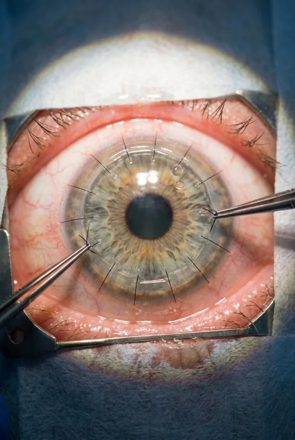
In keratoconus, the cornea gradually thins and bulges forward into a cone shape, causing blurred and distorted vision that glasses correct less and less well. It usually begins in the late teens or early twenties and can worsen over 10–20 years. Risk factors include a family history (about 1 in 10 people with keratoconus have a relative with it), frequent eye rubbing, eye allergies and some connective-tissue disorders. Young people whose glasses prescription keeps changing should ask to be checked for it.
Keratitis means inflammation of the cornea; a corneal ulcer is an open sore on it. Most ulcers are caused by infection, by bacteria, viruses such as herpes simplex, fungi or the Acanthamoeba parasite, but injuries, burns, severe dry eye and eyelids that do not close properly can also cause them. Contact lens wear, especially sleeping in lenses, is a major risk factor. A corneal ulcer is an emergency: untreated, it can permanently damage vision and even cause blindness.
Dystrophies are usually inherited conditions in which material builds up or cells fail in one layer of the cornea, often in both eyes. One of the best known is Fuchs' endothelial dystrophy: the endothelial cells gradually fail, the cornea swells, and vision is typically blurry in the morning (the cornea swells overnight with the eyelids closed), with glare and halos around lights. It rarely shows before the age of 30–40 and was the single most common reason for corneal transplantation in the United States in 2014. Other dystrophies, such as map-dot-fingerprint and lattice dystrophy, affect other layers; those in the front layers can often be treated with laser (PTK).
Scars form when an infection, injury, surgery or inflammation damages Bowman's layer or the stroma. A scar in the centre blurs vision or causes glare; one at the edge may cause no symptoms. How a scar is treated depends on its depth.
A corneal abrasion is a scratch on the surface, often from a fingernail, make-up brush, branch or eye rubbing. It hurts, waters and feels as if something is stuck in the eye, but most heal in 1–2 days, larger ones in about a week. Sometimes the surface layer does not stick down properly afterwards and keeps peeling off again (recurrent corneal erosion), typically causing sudden pain on waking.
Dry eye disease, allergic eye disease, shingles affecting the eye (varicella-zoster) and pterygium (a growth from the white of the eye onto the cornea, more common in sunny climates) can also damage the cornea.
Because the cornea is packed with nerves and must stay perfectly clear, damage usually announces itself. The most common damaged cornea symptoms are:
| Symptom | What it can point to |
|---|---|
| Pain, or a feeling that something is in the eye | Abrasion, foreign body, ulcer, recurrent erosion |
| Redness, watering and sensitivity to light | Keratitis or ulcer, abrasion, inflammation |
| Discharge or a white spot on the cornea | Corneal ulcer: urgent |
| Blurred or hazy vision | Swelling, scarring, infection, dry eye |
| Distorted vision, frequent prescription changes, rising astigmatism | Keratoconus |
| Vision worse in the morning, improving through the day | Fuchs' dystrophy (corneal swelling) |
| Glare and halos around lights | Swelling, scarring, keratoconus, after laser surgery |
| Sudden pain on waking | Recurrent corneal erosion |
Some corneal diseases, especially early keratoconus and dystrophies, cause few symptoms at first and are found on a routine examination or before laser eye surgery.
Examination
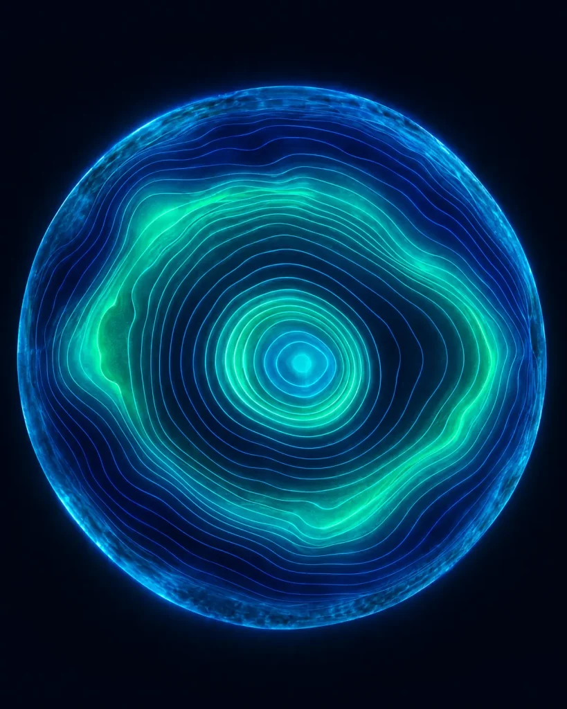
An eye specialist uses several tests, most of them quick and painless:
At Laser Istanbul Clinic, corneal topography, OCT and biometry are part of every pre-operative examination in Istanbul, and your surgeon reviews your existing scans before you book. About corneal topography
| Treatment | What it does | Typically used for |
|---|---|---|
| Medicated eye drops | Antibiotic, antifungal or antiviral drops treat infection; lubricants and, once infection is controlled, steroid drops calm the surface | Ulcers and keratitis, abrasions, dry eye |
| Bandage or special contact lenses | Protect a healing surface, or give a smooth new optical surface over an irregular cornea (rigid gas-permeable and scleral lenses) | Abrasions and recurrent erosion; keratoconus |
| Corneal cross-linking (CXL) | Riboflavin (vitamin B2) drops plus ultraviolet light strengthen and stiffen the corneal tissue; FDA-approved in 2016 and the main treatment that can stop keratoconus getting worse | Progressive keratoconus |
| Corneal ring segments (e.g. Intacs) | Small curved implants placed in the cornea to flatten the cone | Moderate keratoconus |
| Phototherapeutic keratectomy (PTK) | An excimer laser removes a thin, precise layer from the front of the cornea | Superficial scars, front-layer dystrophies, recurrent erosion |
| Laser reshaping (LASIK, SMILE, PRK) | Reshapes the stroma to change the cornea's focusing power | Short-sightedness, long-sightedness and astigmatism in healthy corneas only |
| Corneal transplant (keratoplasty) | Replaces damaged corneal tissue with healthy donor tissue | Advanced keratoconus, deep scars, endothelial failure, severe infection |
A corneal transplant uses tissue from a deceased donor, checked to make sure it is healthy before use. Surgeons now replace only the layers that are damaged whenever they can:
| Type | Layers replaced | Typically used for | Recovery (NHS guidance) |
|---|---|---|---|
| PK (penetrating keratoplasty) | Full thickness, held with stitches | Damage through all layers | Up to 2 years |
| DALK (deep anterior lamellar keratoplasty) | Front and middle layers; your own endothelium is kept | Keratoconus and scars when the endothelium is healthy | Up to 18 months |
| DSAEK (Descemet's stripping automated endothelial keratoplasty) | Endothelium, Descemet's membrane and a thin layer of back stroma | Fuchs' dystrophy, corneal swelling | Up to 6 months |
| DMEK (Descemet's membrane endothelial keratoplasty) | Only Descemet's membrane and endothelium, held in place by an air or gas bubble | Fuchs' dystrophy, corneal swelling | Up to 6 months |
Good to know
DMEK uses the thinnest graft and a smaller incision than DSAEK (about 2.8 mm versus 5 mm). In one comparison, average corrected vision a year after surgery was 20/24 with DMEK and 20/32 with DSAEK, and DMEK has a lower rejection rate. The pre-Descemet's layer is what makes the "big bubble" technique used in many DALK operations possible.
Rejection can happen at any time after a transplant and can usually be treated with steroid drops if it is caught quickly. Warning signs include a red eye, pain, sensitivity to light and blurred vision.
Can laser eye surgery be done on any cornea? No. The cornea has to be healthy and thick enough. The American Academy of Ophthalmology lists corneas that are too thin, corneal scars or disease, keratoconus and severe dry eye among the reasons a person is not a candidate for LASIK. This is why topography and pachymetry are always measured first.
Cornea and keratoconus care, cross-linking and corneal transplants (DMEK, DSAEK, DALK and full-thickness grafts) are led by Doç. Dr. Muhammet Derda Özer, whose published research includes corneal cross-linking for keratoconus in young people.
If you already have a diagnosis, such as keratoconus or Fuchs' dystrophy, and want a second opinion on cross-linking or a transplant, you can send your scans for review before you travel.
The cornea is the clear, dome-shaped front surface of the eye. It covers the iris and pupil, protects the eye and provides about two-thirds of its focusing power.
From front to back: the epithelium, Bowman's layer, the stroma, Descemet's membrane and the endothelium. A sixth, very thin pre-Descemet's (Dua's) layer between the stroma and Descemet's membrane was described in 2013.
It depends on the layer. The surface epithelium heals within days, but damage to Bowman's layer or the stroma heals with scar tissue, and lost endothelial cells are not replaced. Deep scars or endothelial failure may need laser treatment or a transplant.
Yes. A corneal transplant replaces damaged corneal tissue with healthy donor tissue. Today surgeons often replace only the diseased layers (DALK, DSAEK or DMEK) rather than the full thickness (PK), which usually means faster recovery.
Laser eye surgery deliberately reshapes the corneal stroma to change its focusing power. In a healthy, thick enough cornea this is safe for most people, but it cuts some corneal nerves, which often causes temporary dry eye. That is why the cornea is measured in detail before any laser treatment.
Because it has no blood vessels, its collagen fibres are arranged in a precise, regular pattern, and its inner endothelial layer constantly pumps out excess water.
Send us your scans or eye report. Doç. Dr. Muhammet Derda Özer, our cornea specialist, will review them and tell you which treatments may suit your eyes, with no obligation.
Your case is reviewed by a named surgeon, not a salesperson. Reply within {{response time}}.
 Prof. Dr. Ahmet Satıcı
 Doç. Dr. Muhammet Derda Özer
 Op. Dr. Aysan Bayram