
- Home: eye surgery in Istanbul
- All eye treatments
- Retina surgery: vitrectomy & retinal detachment
Vitrectomy & Retina Surgery in Turkey: Retinal Detachment, Macular Hole and Retinal Laser in Istanbul
Vitrectomy is the keyhole operation at the heart of modern eye surgery on the retina: it repairs detached retinas, closes macular holes, peels scar tissue off the macula and clears bleeding inside the eye. In Istanbul, your retina care is led by Doç. Dr. Muhammet Derda Özer (Doç. = Associate Professor) or Op. Dr. Aysan Bayram, who examine your retina, explain which treatment suits your eye and stay with you through recovery, with a personal interpreter at every appointment.
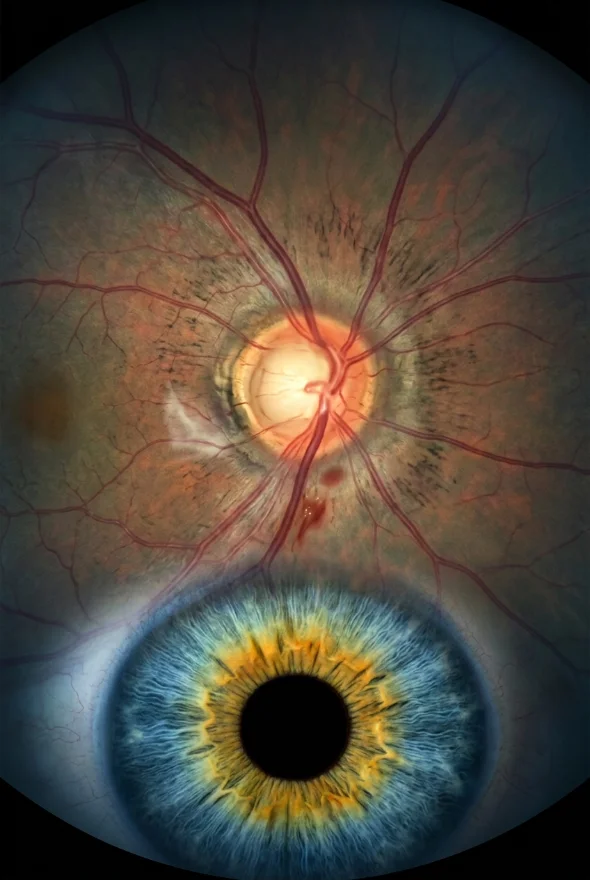
Vitrectomy removes the clear gel (vitreous) from inside the eye so a retina surgeon can repair the retina directly, usually through three openings smaller than 1 mm, then fills the eye with saline, gas or silicone oil. It treats retinal detachment, macular holes, macular pucker and diabetic bleeding. In Istanbul it is performed by Doç. Dr. Özer or Op. Dr. Bayram from {{£ price}} per eye; hotel and transfers are extra.
Sudden flashes, a shower of new floaters or a dark curtain? Get seen today.
The NHS, the American Academy of Ophthalmology (AAO) and the US National Eye Institute (NEI) all give the same advice: new floaters or flashes, a rapid increase in them, a shadow or "curtain" moving across your vision, or sudden blurring need an eye doctor straight away, because they can be the first signs of a retinal tear or detachment. Do not wait to travel for treatment. If you are already in Turkey, message us and we will arrange an urgent retina examination {{urgent appointment availability, e.g. "the same day"}}. If you are at home, use your local emergency eye service first; we are happy to review your scans afterwards.
Retina anatomy, and how retinal vision works
The retina is a thin layer of nerve tissue, about 0.5 mm thick, that lines the back of the eye and turns light into signals for the brain. It works like the sensor in a camera: the cornea and lens focus light onto it, light-sensing cells (rods and cones) convert it into electrical signals, and those signals leave the eye through the optic nerve. According to the University of Pittsburgh's Webvision retina textbook, light has to pass through the full thickness of the retina before it reaches the rods and cones, which sit at the very back against the pigment layer.
Retinal vision has two jobs, and different parts of the retina do them:
- The macula and its centre, the fovea, give sharp central vision for reading, faces and colour. The fovea is less than a quarter of a millimetre across and contains only cones, packed tightly together.
- The peripheral retina is dominated by rods. It gives side vision and vision in dim light, and it is where most retinal tears start.
This is why a retinal problem can feel so different from person to person: a small hole in the macula can make reading impossible while side vision stays normal, whereas a detachment that starts at the edge may first show as a shadow at the side.
In front of the retina sits the vitreous, a clear gel that fills the back of the eye. With age it shrinks and pulls away from the retina (posterior vitreous detachment). That is normal, but when it tugs too hard it can tear the retina or pull on the macula, and many of the operations on this page start by removing it.
The 10 layers of the retina, in order
Under a microscope, and on an OCT scan, the retina shows ten layers. Listed from the back of the eye (outermost) to the front (innermost, facing the vitreous):
| # | Layer | What it contains or does |
|---|---|---|
| 1 | Retinal pigment epithelium (RPE) | A single layer of pigmented cells that supports and nourishes the rods and cones |
| 2 | Photoreceptor layer | The light-sensing outer parts of the rods and cones |
| 3 | Outer (external) limiting membrane | Junctions between Müller support cells and the photoreceptors |
| 4 | Outer nuclear layer | Cell bodies of the rods and cones |
| 5 | Outer plexiform layer | Connections between photoreceptors and bipolar and horizontal cells |
| 6 | Inner nuclear layer | Cell bodies of bipolar, horizontal and amacrine cells |
| 7 | Inner plexiform layer | Connections between bipolar cells and ganglion cells |
| 8 | Ganglion cell layer | Cell bodies of ganglion cells, the output neurons of the retina |
| 9 | Nerve fibre layer | Ganglion cell axons running to the optic nerve |
| 10 | Inner limiting membrane (ILM) | The end feet of Müller cells and a basement membrane, facing the vitreous |
Strictly speaking, the RPE is the outer wall the nerve retina rests on, and a retinal detachment is a separation between layers 1 and 2. The innermost layer matters in surgery too: in macular hole operations the surgeon peels the inner limiting membrane around the hole to help it close.
Layers of the retina diagram
Your own retina can be seen layer by layer on an OCT scan, a quick, non-contact scan that the AAO's EyeWiki describes as a non-invasive cross-sectional view of the living retina. It is how macular holes, epiretinal membranes, macular swelling and detachments are confirmed and measured.
Retinal diseases list: what retina surgeons treat
These are the retinal conditions people most often ask us about, with the usual treatment and where to read more:
| Condition | What happens | Usual treatment |
|---|---|---|
| Retinal tear | The shrinking vitreous pulls a hole or flap in the retina | Retinal laser or freezing, often the same day |
| Retinal detachment | Fluid lifts the retina off the back wall of the eye | Surgery: vitrectomy, scleral buckle or gas injection |
| Macular hole | A small gap opens in the centre of the macula | Vitrectomy with ILM peel and a gas bubble |
| Epiretinal membrane (macular pucker) | A thin film of scar tissue wrinkles the macula | Observation, or vitrectomy with membrane peel if vision is affected |
| Diabetic retinopathy | Damaged and abnormal blood vessels leak, bleed or scar | Laser, anti-VEGF injections, vitrectomy for bleeding or traction |
| Vitreous haemorrhage | Bleeding into the vitreous gel | Wait and monitor, laser, injections or vitrectomy, depending on the cause |
| Age-related macular degeneration (AMD) | The macula wears out (dry) or leaks (wet) | Injections for wet AMD; low-vision aids or a macula lens for late disease |
| Retinal vein occlusion | A blocked retinal vein causes swelling and bleeding | Injections, sometimes laser |
| Lattice degeneration | Thin, weak patches at the edge of the retina | Usually monitoring; laser in selected cases |
| Retinal coloboma | A gap in the retina and choroid present from birth | Monitoring; detachments need specialist surgery |
| Endophthalmitis | Infection inside the eye | Urgent antibiotic treatment; vitrectomy in some cases |
What is retinal coloboma?
A retinal (chorioretinal) coloboma is a gap in the retina and the layer beneath it, present from birth, because a seam in the developing eye did not close in the second month of pregnancy. EyeWiki (AAO) reports that ocular coloboma occurs in about 0.5–2.2 per 10,000 births. How well the eye sees depends mainly on whether the fovea, the centre of the macula, has formed normally. The most important risk is retinal detachment, which EyeWiki reports in 23–42% of people with a coloboma at the back of the eye. Laser around the coloboma may reduce that risk, although no randomised trial has proven it. If you have a coloboma, regular retina checks and knowing the warning signs of detachment matter more than for most people.
What is vitrectomy, and how does it work?
Vitrectomy (pars plana vitrectomy, PPV) is keyhole surgery inside the eye in which the vitreous gel is removed and replaced, so the surgeon can work directly on the retina. The AAO describes it as surgery for problems of the retina and vitreous, in which the gel is replaced with a saline solution, a gas bubble or silicone oil.
How a vitrectomy is done, step by step:
- Numbing. Most operations are done under local anaesthetic, with an injection around the eye; you are awake but feel no pain. General anaesthetic is possible.
- Three tiny openings. The surgeon places three ports in the white of the eye, in a zone called the pars plana, for a light, a fluid line and the cutting instrument. Modern ports are 23-, 25- or 27-gauge, about 0.6, 0.5 and 0.4 mm across, and usually need no stitches.
- Removing the gel. A fine cutter removes the vitreous, together with any blood, scar tissue or traction pulling on the retina.
- Repairing the retina. Depending on the problem, the surgeon flattens a detached retina, peels a membrane off the macula, closes a macular hole or treats tears with laser applied from inside the eye (endolaser) or freezing.
- Filling the eye. The eye is filled with saline, air, a longer-lasting gas, or silicone oil, which holds the retina in place while it heals.
How long does vitrectomy take? The AAO says from one to several hours, depending on what needs to be repaired; NHS information gives about one hour for a typical macular hole operation.
The bubble decides your trip. Gas bubbles are absorbed on their own; NHS patient leaflets give up to about 2 weeks for air, 4 weeks for SF6, 8 weeks for C2F6 and 12 weeks for C3F8. You cannot fly, travel to high altitude or have nitrous oxide anaesthetic while gas is in the eye, because the bubble expands and can raise eye pressure enough to cause severe pain and permanent sight loss. Silicone oil does not disappear, so it is usually removed in a second, smaller operation a few months later.
A little history. EyeWiki (AAO) credits Robert Machemer with the first closed vitrectomy in 1971. Instruments have since shrunk from 17-gauge to today's sutureless micro-incision systems, which EyeWiki links with more comfort and less change to the shape of the cornea.
Technology we use: {{vitrectomy system name and gauge, e.g. "23/25-gauge"}}, a wide-angle viewing system, endolaser and {{tamponade options available: air / SF6 / C2F6 / C3F8 / silicone oil}}, in a fully equipped, Ministry of Health–licensed operating theatre in Istanbul.
Who needs vitrectomy?
You may need vitrectomy if the vitreous gel is pulling on, bleeding into or blocking the view of your retina, or if the retina itself needs repairing from the inside. The AAO lists these reasons:
- retinal detachment, especially after cataract surgery, with large or multiple tears, or with bleeding inside the eye
- macular hole
- macular pucker (epiretinal membrane) that distorts or blurs vision
- diabetic retinopathy with bleeding or scar tissue pulling on the retina
- infection inside the eye (endophthalmitis)
- severe eye injury
- complications of cataract surgery, such as a piece of lens dropped into the back of the eye
When vitrectomy is usually not the first choice: a retinal tear that has not detached is normally sealed with laser alone; wet macular degeneration is treated with injections, not surgery; and ordinary floaters usually need no treatment at all, as the NHS notes. A retinal detachment in a younger eye with its natural lens may be better repaired with a scleral buckle, and some single upper tears with a gas injection alone.
Who should not travel for surgery? Anyone whose detachment threatens the macula and who is not already in Istanbul should be treated at home, without delay. Travel makes sense for planned surgery (macular hole, epiretinal membrane, longstanding diabetic bleeding), a second opinion, follow-on surgery such as silicone-oil removal, or care while you are already in Turkey.
How successful is retina surgery? Published results by condition
| Condition | Operation | What published studies report |
|---|---|---|
| Retinal detachment | Vitrectomy, scleral buckle or gas injection | NHS consent information: an 85–90% chance that one operation reattaches the retina and it stays attached. In the PIVOT trial (2019), vitrectomy succeeded with one operation in 93% of eyes. |
| Macular hole | Vitrectomy with ILM peel and gas | A 2020 literature review reports closure in more than 90% of holes with ILM peeling, even large holes up to 650 µm. |
| Epiretinal membrane | Vitrectomy with membrane peel | EyeWiki (AAO): average vision improved from about 20/110 to 20/55 in a meta-analysis; most improvement comes within 3–6 months, and the membrane returns in about 1% of eyes. |
| Vitreous haemorrhage | Vitrectomy | A 2023 review calls vitrectomy the cornerstone of treatment when blood does not clear; the outcome depends on the cause. |
What "success" means. A closed hole or a flat retina does not always mean perfect sight. How much vision returns depends on how long the problem was present and whether the macula was involved, which is why your surgeon gives you a personal estimate after your OCT scan rather than a single percentage.
Retinal laser: types of retinal lasers and their side effects
A retinal laser uses focused light to make tiny, controlled scars on the retina, either to weld a tear down or to calm abnormal blood vessels. It is done at the slit-lamp microscope with numbing drops, usually in about 20 minutes, without any cut. The two main uses are laser retinopexy around a tear and panretinal photocoagulation (PRP) for diabetic eye disease.
Types of retinal lasers
| Type | What it is | Typical use |
|---|---|---|
| Argon laser (488 nm blue / 514 nm green) | The original retinal laser; "argon laser" is still the name most patients use | Retinal tears, diabetic retinopathy |
| 532 nm green laser (frequency-doubled Nd:YAG) | The modern successor that does the same job; one of the most widely used retinal lasers today | Retinal tears, PRP, some vein occlusions |
| 810 nm diode laser | Infrared laser, also widely used today | Retinal photocoagulation |
| Pattern-scan laser | Fires a grid of short spots (about 20 milliseconds instead of the classic 100–200) | PRP with less heat spread and more comfort |
| Endolaser | Laser delivered through a probe inside the eye during vitrectomy | Sealing tears and treating diabetic retina during surgery |
| Photodynamic therapy (PDT) | A light-activated drug with a low-energy laser | Selected wet AMD and other macular conditions; no longer first-line for AMD |
Side effects of retinal tear laser surgery
Retinal tear laser (laser retinopexy) is usually well tolerated, and because there is no incision, NHS patient leaflets note there is no risk of infection inside the eye. Common, short-lived effects are:
- discomfort, or brief sharp or aching sensations, during treatment, and sometimes a headache afterwards
- dazzled or misty vision for a few hours; no driving on the day
- a red eye that settles within a few days
- floaters that may seem to increase for a few days; laser does not remove floaters
Less often, the tear does not fully seal and needs more laser, bleeding occurs, or a thin film (epiretinal membrane) forms on the retina. A detachment can still happen after laser, but NHS patient information puts the risk at less than 1 in 20 with treatment, compared with about half of untreated symptomatic tears. The scar takes about two weeks to become strong.
With PRP for diabetic retinopathy, EyeWiki (AAO) notes that some loss of peripheral, colour and night vision can be permanent, which your surgeon weighs against the much larger risk of sight loss from untreated disease.
Eye dilation vs retinal imaging: do you still need the drops?
Retinal imaging is a useful record, but it does not replace a dilated retinal examination when a tear or detachment is suspected. The NEI calls the dilated eye exam the only way to check for many eye diseases early, before they cause vision loss. A 2025 review of retinal detachment explains that the key examination is still a dilated look at the retina with an indirect ophthalmoscope, often with gentle pressure on the eye (scleral indentation) to see the far edge of the retina, where tears hide. Ultra-widefield photographs help document a detachment and help patients, such as children, who cannot tolerate that examination.
| Dilated retinal examination | Retinal imaging (widefield photo, OCT) | |
|---|---|---|
| What it shows | The whole retina in 3D, including the far edge with indentation | A photo of most of the retina (widefield) or a cross-section of the macula (OCT) |
| Drops needed | Yes; vision is blurred and light-sensitive for a few hours (NEI) | Often possible without drops, though some scans are clearer with them |
| Best for | Finding retinal tears, lattice and detachments | Recording the retina, measuring the macula, tracking change over time |
| Limits | Takes longer; no driving until the drops wear off | Can miss small tears at the very edge of the retina |
Before any retina surgery with us you have both: a dilated examination by your surgeon and the scans (OCT, and ultrasound if blood blocks the view). Bring someone with you or plan not to drive after the drops.

Can a damaged retina repair itself?
Mostly not: lost retinal nerve cells do not grow back in humans, so the aim of treatment is to protect what is left. Researchers studying retinal regeneration note that fish such as zebrafish can regrow retinal neurons, but in mammals the retina's support cells (Müller glia) respond to damage by forming scar-like tissue instead, which can lead to further cell loss and permanent vision loss.
What can recover:
- A reattached retina often regains function slowly. The AAO says sight usually begins to improve four to six weeks after detachment surgery, and the retina can continue healing for a year or more.
- Swelling of the macula from diabetes, vein blockage or wet AMD can shrink with injections, and vision can improve when it does.
- A closed macular hole often brings back some central vision.
What does not recover: retinal cells that have died, laser scars, and long-standing damage. Stem cell treatments for the retina are still experimental; the AAO says no stem cell product is approved for eye disease in the US. This is why speed matters with retinal symptoms.
Retina surgery in Turkey: price and what is included
At Laser Istanbul Clinic, vitrectomy starts from {{£ price}} per eye. The final price depends on the operation (detachment repair, macular hole, membrane peel, diabetic surgery), whether silicone oil or combined cataract surgery is needed, and how complex the eye is. Your surgeon confirms it in writing after reviewing your scans, before you book anything.
| Retina treatment with us | From |
|---|---|
| Retina assessment with dilated examination and OCT | {{£ price}} |
| Vitrectomy (macular hole, epiretinal membrane, vitreous haemorrhage), per eye | {{£ price}} |
| Retinal detachment surgery, per eye | {{£ price}} |
| Silicone-oil removal, per eye | {{£ price}} |
| Retinal laser (argon / 532 nm), per eye | {{£ price}} |
| Anti-VEGF injection, per injection | {{£ price}} |
Typical published prices for vitrectomy, per eye (2025–26):
| Where | Typical price range |
|---|---|
| UK, private | about $8,000–13,400 |
| USA | about $8,000–14,000 |
| Istanbul (published package prices) | about $3,200–6,500 |
| Istanbul with Laser Istanbul Clinic | from {{£ price}} |
Typical ranges advertised by clinics and price-comparison sites in 2025–26, not quotes. Published Istanbul prices are often packages that include a hotel. In the UK, an acute retinal detachment is treated urgently through the NHS.
Your treatment price includes:
- full retina examination and scans (including OCT) with your surgeon
- surgery by your named retina surgeon
- medication and eye drops for your recovery
- follow-up checks in Istanbul with the same surgeon
- a personal interpreter, a dedicated coordinator and remote follow-up after you return home
Not included: flights, hotel and airport transfers. Hotel and transfers are a separate charge, and we can arrange them for you if you ask. If your eye needs a gas bubble, you cannot fly until it has gone, so plan and budget for a longer stay.
Your retina surgeons



Your retina surgery journey, step by step
- Send your scans
Upload your eye report, OCT or ultrasound images and a description of your symptoms. A retina surgeon replies within {{response time}}. If your symptoms are new, we will tell you to get seen locally first.
- Plan your dates
For planned vitrectomy, allow {{typical stay}} in Istanbul. If a gas bubble is likely, plan to stay until you are cleared to fly (2–12 weeks depending on the gas), or ask your surgeon whether another option suits you.
- Retina examination (day 1)
Dilated examination, OCT scan, ultrasound if needed, eye pressure and a check of the other eye. Your surgeon explains the operation, the expected result and the risks for your eye.
- Surgery
Usually under local anaesthetic, in a fully equipped, Ministry of Health–licensed operating theatre in Istanbul. You go back to your hotel the same day with a pad and shield over the eye.
- Positioning, if advised
With gas or oil, you may need to keep your head face-down or to one side for part of each day, for up to about 7 days. Your surgeon tells you exactly how.
- Checks in Istanbul
The next day and again before you leave; your surgeon confirms when you can fly.
- Home and remote follow-up
Your coordinator stays in touch, your surgeon reviews your progress by video, and we send a written report for your eye doctor at home.
Recovery after vitrectomy: what to expect
-
First days
Eye padded and shielded; a scratchy, sandy feeling is common (AAO). Vision is very blurred while a gas bubble fills the eye.
-
First 1–2 weeks
Positioning if advised. Drops exactly as prescribed; shield at night. No rubbing, swimming or eye make-up.
-
2–6 weeks
The eye heals and redness settles. As gas absorbs, you see a wobbling line, like a spirit level, that moves down until the bubble disappears.
-
2–12 weeks
Gas fully absorbed, depending on the type. Only then is flying or high-altitude travel safe.
-
3–6 months
Most improvement after epiretinal membrane surgery; vision after macular hole and detachment surgery keeps improving. A new glasses prescription may help.
-
Up to a year or more
The retina can continue to recover. Silicone oil, if used, is usually removed in this period.
Reading, television and screens do not harm the eye. Do not drive until your surgeon confirms your vision meets the driving standard. Call your coordinator straight away if you have severe pain, increasing redness or a sudden drop in vision.
Risks and side effects of vitrectomy
Vitrectomy is a well-established operation, but it is surgery inside the eye, and you should know the risks before you decide.
Cataract is the most predictable effect. If you still have your natural lens, NHS information on macular hole surgery says a cataract is almost certain within a year, and a 2025 review notes that most eyes need cataract surgery within two years of a vitrectomy. The AAO adds that this is especially likely after age 50. Cataract is treated with routine cataract surgery; some patients have both done together.
Other possible complications listed by the AAO and EyeWiki:
- a new retinal tear or detachment
- raised pressure inside the eye, which can lead to glaucoma if it persists
- bleeding inside the eye
- swelling of the macula
- infection inside the eye (endophthalmitis): rare, but serious
- poor vision, or the problem not being fully fixed
- for detachments, scar tissue (proliferative vitreoretinopathy) that can pull the retina off again; NHS guidance puts the chance of further surgery at 5–10%
Gas and altitude. Flying, mountain travel, scuba diving or nitrous oxide anaesthetic while gas is in the eye can cause a dangerous rise in eye pressure. Many surgeons give you a wristband to show any doctor.
Your surgeon explains your personal risks in writing and through your interpreter before you sign anything.
Vitrectomy and retina surgery: frequently asked questions
Vitrectomy is keyhole eye surgery that removes the clear gel inside the eye (the vitreous) so the surgeon can repair the retina from inside. The gel is replaced with saline, gas or silicone oil. It is used for retinal detachment, macular holes, macular pucker, diabetic bleeding and some eye injuries and infections.
The AAO says it takes from one to several hours, depending on the repair; a typical macular hole operation takes about an hour. Most patients have local anaesthetic, so they are awake, but the eye is numb and they feel no pain. General anaesthetic is possible if needed.
The eye usually takes 2 to 6 weeks to heal, and vision keeps improving for months. You cannot fly while gas is in the eye: NHS patient leaflets give about 2 weeks for air, 4 weeks for SF6, 8 weeks for C2F6 and 12 weeks for C3F8. With saline or silicone oil, your surgeon tells you when flying is safe.
Probably, if you still have your natural lens. A cataract develops in most eyes after vitrectomy, often within one to two years, especially after age 50. It is treated with routine cataract surgery, which our surgeons can plan with you.
From the back of the eye forwards: retinal pigment epithelium, photoreceptor layer (rods and cones), outer limiting membrane, outer nuclear layer, outer plexiform layer, inner nuclear layer, inner plexiform layer, ganglion cell layer, nerve fibre layer and inner limiting membrane. OCT scans show these layers in the living eye.
Not fully. Retinal nerve cells that die do not grow back in humans, but a reattached retina can regain function over months, and swelling of the macula can settle with treatment. The sooner a retinal problem is treated, the more vision is saved.
Usually mild and short-lived: discomfort during treatment, a headache, dazzled vision for a few hours, a red eye for a few days and a brief increase in floaters. Less often, the tear needs more laser, or bleeding or a thin membrane develops. The laser greatly lowers, but does not remove, the risk of detachment.
A retinal coloboma is a gap in the retina and the layer beneath it, present from birth, because part of the developing eye did not close. Vision depends on whether the centre of the retina is affected. People with a coloboma at the back of the eye have a high risk of retinal detachment, reported at 23–42%, so regular retina checks are important.
If a retinal tear or detachment is possible, you need a dilated examination: it is the best way to see the far edge of the retina. Retinal photos and OCT scans add a record and measure the macula, but they can miss small tears at the edge. Most retina assessments use both.
Published prices for vitrectomy in Istanbul are about $3,200–6,500 per eye, compared with about $8,000–14,000 privately in the UK or USA. With Laser Istanbul Clinic it starts from {{£ price}} per eye, including examination and scans, surgery, medication, follow-up checks, interpreter and coordinator. Hotel and transfers are extra.
Retina care is led by Doç. Dr. Muhammet Derda Özer, an Associate Professor of Ophthalmology whose work covers medical and surgical retina, and Op. Dr. Aysan Bayram, whose focus includes retinal disease. You know your surgeon's name before you book.
Retina treatments in Istanbul, and how they differ
| Vitrectomy | Retinal detachment surgery | Argon laser | Intravitreal injections | Macular degeneration care | |
|---|---|---|---|---|---|
| Treats | Macular hole, macular pucker, diabetic bleeding, detachment | Detached retina | Retinal tears, lattice, diabetic retinopathy | Wet AMD, diabetic macular oedema, vein occlusion | Wet and dry AMD |
| Where | Operating theatre | Operating theatre | Clinic, about 20 minutes | Clinic, minutes | Clinic, or theatre for a macula lens |
| Stay in Istanbul | Longer if a gas bubble is used | Longer if a gas bubble is used | Short | Repeated visits over months | Depends on the treatment |
Vitrectomy
Keyhole retina surgery for macular holes, epiretinal membranes, bleeding and detachments.
Vitrectomy in IstanbulRetinal detachment surgery
Vitrectomy, scleral buckle or gas injection, with clear rules on positioning and flying.
Retinal detachment surgeryArgon laser
Seals retinal tears before they detach and treats diabetic retinopathy.
Argon laser (retinal laser photocoagulation)Anti-VEGF eye injections
For wet macular degeneration, diabetic macular swelling and vein occlusion.
Intravitreal injections (anti-VEGF eye injections)Macular degeneration
Injections for wet AMD and the Scharioth macula lens for reading in late AMD.
Macular degeneration treatment & Scharioth macula lensOther eye treatments in Istanbul
Many retina patients also ask about lens and pressure problems:
Cataract surgery in IstanbulRetina and glaucoma guides
- Retinal detachment: warning signs and urgent surgery (/en/eye-health/retina-glaucoma/retinal-detachment-warning-signs-and-urgent-surgery/)
- Eye floaters: when to worry and when vitrectomy helps (/en/eye-health/retina-glaucoma/eye-floaters-when-to-worry-and-when-vitrectomy-helps/)
- Glaucoma: symptoms, causes and when surgery is needed (/en/eye-health/retina-glaucoma/glaucoma-symptoms-causes-and-when-surgery-is-needed/)
- Glaucoma surgery options: SLT, MIGS, trabeculectomy (/en/eye-health/retina-glaucoma/glaucoma-surgery-options-slt-migs-trabeculectomy/)
- Trabeculectomy vs trabeculotomy vs MIGS: which glaucoma operation? (/en/eye-health/retina-glaucoma/trabeculectomy-vs-trabeculotomy-vs-migs/)
Sources for the facts on this page
- American Academy of Ophthalmology, What Is Vitrectomy?: definition; gel replaced with saline, gas bubble or silicone oil; indications (diabetic retinopathy with bleeding or scar tissue, retinal detachment, macular hole, macular pucker, endophthalmitis, severe injury, cataract-surgery complications); local or general anaesthetic; one to several hours; eye patch, scratchy feeling; face-down or side positioning with gas; no flying, mountains or scuba diving with gas; risks (infection, bleeding, torn or detached retina, poor vision, raised pressure/glaucoma, cataract especially over 50). Opened 6 Oct 2026. https://www.aao.org/eye-health/treatments/what-is-vitrectomy
- EyeWiki (AAO), Pars Plana Vitrectomy: indications; 20/23/25/27-gauge = 0.9/0.6/0.5/0.4 mm; smaller gauges with more comfort and less corneal astigmatism; Machemer closed vitrectomy 1971, 3-port 20-gauge 1974; SF6 and C3F8 durations; complications (cataract, glaucoma, retinal detachment, hypotony, cystoid macular oedema). https://eyewiki.org/Pars_Plana_Vitrectomy
- NHS, Macular hole: definition and symptoms; vitrectomy with ILM peel and gas bubble; local or general anaesthetic, most choose local; about one hour; face-down positioning; no flying or high altitude with gas (up to 12 weeks); cataract almost certain within a year; other risks. https://www.nhs.uk/conditions/macular-hole/
- National Eye Institute (NIH), Macular Hole: vitrectomy with gas bubble as a temporary bandage; avoid flying and nitrous oxide until the bubble is gone. https://www.nei.nih.gov/learn-about-eye-health/eye-conditions-and-diseases/macular-hole
- EyeWiki (AAO), Macular Hole: incidence 8.69 per 100,000/year; positioning data (94% vs 84% closure for holes ≥400 µm, Cochrane). https://eyewiki.org/Macular_Hole
- Ittarat M, Somkijrungroj T, Chansangpetch S, Pongsachareonnont P. Literature Review of Surgical Treatment in Idiopathic Full-Thickness Macular Hole. Clin Ophthalmol 2020 (PMC7398756): anatomical closure > 90% with ILM peeling, even in holes up to 650 µm. https://pmc.ncbi.nlm.nih.gov/articles/PMC7398756/
- EyeWiki (AAO), Epiretinal Membrane: prevalence 7–11%; surgery for significant visual decline or distortion; mean VA 20/110 → 20/55 (meta-analysis); improvement within 3–6 months, up to 1–2 years; recurrence ~1%; complications incl. cataract, retinal breaks and detachment. https://eyewiki.org/Epiretinal_Membrane
- Shaikh N, et al. Vitreous hemorrhage – Causes, diagnosis, and management. Indian J Ophthalmol 2023 (PMC10155538): treatment options; pars plana vitrectomy the cornerstone of management; prognosis depends on cause. https://pmc.ncbi.nlm.nih.gov/articles/PMC10155538/
- Xiong J, Tran T, Waldstein SM, Fung AT. A review of rhegmatogenous retinal detachment: past, present and future. Wien Med Wochenschr 2025 (PMC12031774): dilated indirect ophthalmoscopy with scleral depression to the ora serrata as the key examination; ultra-widefield photography helpful for documentation and patients who cannot tolerate indentation; OCT non-invasive; micro-incisional 23–27-gauge sutureless vitrectomy since the 2000s; most eyes need cataract extraction within 2 years of vitrectomy; postoperative complications list. https://pmc.ncbi.nlm.nih.gov/articles/PMC12031774/
- Webvision (University of Pittsburgh), Kolb H, Simple Anatomy of the Retina: retina ~0.5 mm thick, lines the back of the eye; light passes through the retina to the rods and cones, which lie against the pigment epithelium; fovea < 0.25 mm (200 µm), cone-only; central retina cone-dominated, peripheral rod-dominated; contents of the outer/inner nuclear and plexiform layers, ganglion cell and nerve fibre layers; outer and inner limiting membranes formed by Müller cells; optic nerve = ganglion cell axons. https://www.webvision.pitt.edu/book/part-i-foundations/simple-anatomy-of-the-retina/ (and Photoreceptors: fovea contains only cones. https://www.webvision.pitt.edu/book/part-ii-anatomy-and-physiology-of-the-retina/photoreceptors/)
- EyeWiki (AAO), Optical Coherence Tomography: non-invasive cross-sectional view of the retina; retinal layers on OCT (ILM to RPE/Bruch complex); diagnoses macular holes, epiretinal membranes, macular oedema, detachments, AMD. https://eyewiki.org/Optical_Coherence_Tomography
- EyeWiki (AAO), Coloboma: failed closure of the embryonic fissure in the second month of gestation; 0.5–2.2 per 10,000 live births; foveal anatomy determines vision; retinal detachment in 23–42% of posterior colobomas; prophylactic laser may reduce detachment (no randomised trial). https://eyewiki.org/Coloboma
- National Eye Institute (NIH), Get a Dilated Eye Exam: the only way to check for many eye diseases early; blurry, light-sensitive vision for a few hours afterwards. https://www.nei.nih.gov/learn-about-eye-health/healthy-vision/get-dilated-eye-exam
- Campbell LJ, Levendusky JL, Steines SA, Hyde DR. Retinal regeneration requires dynamic Notch signaling. Neural Regen Res 2022 (PMC8643038): zebrafish Müller glia regenerate retinal neurons; mammalian Müller glia enter a prolonged gliotic state leading to further neuronal death and permanent vision loss. https://pmc.ncbi.nlm.nih.gov/articles/PMC8643038/ (see also Ramachandran R et al., Nat Cell Biol 2010, PMC2972404: "unlike mammals", fish mount a robust regenerative response.)
- American Academy of Ophthalmology, Stem Cell Therapy for Eye Disease: What You Need to Know (4 Jun 2019): no FDA-approved stem cell products for eye disease. https://www.aao.org/eye-health/tips-prevention/stem-cell-therapy-eye-disease-facts
- NHS, Floaters and flashes in the eyes: treatment usually needed only if a problem could affect vision; urgent signs. https://www.nhs.uk/conditions/floaters-and-flashes-in-the-eyes/
- NHS, Detached retina (retinal detachment): symptoms, urgency, treatments, recurrence. https://www.nhs.uk/conditions/detached-retina-retinal-detachment/
- NHS patient leaflet, Retinal detachment surgery (Moorfields Eye Hospital NHS Foundation Trust): 85–90% one-operation success; 5–10% further surgery; gas durations (air up to 2 weeks, SF6 4, C2F6 8, C3F8 12); silicone oil removed later; nitrous oxide warning; openings < 1 mm; healing 2–6 weeks; positioning up to 7 days; reading/TV safe. https://www.moorfields.nhs.uk/mediaLocal/s1jdmews/retinal-detachment-surgery-_0.pdf
- American Academy of Ophthalmology, Detached Retina: sight improves 4–6 weeks after surgery; retina heals for a year or more; no flying, altitude or scuba diving with gas. https://www.aao.org/eye-health/diseases/detached-torn-retina
- Hillier RJ et al. PIVOT trial. Ophthalmology 2019;126:531–539: primary success 93.2% vitrectomy vs 80.8% pneumatic retinopexy. https://www.aaojournal.org/article/S0161-6420(18)32275-9/fulltext
- EyeWiki (AAO), Lasers (surgery): argon 488/514 nm; 532 nm frequency-doubled Nd:YAG and 810 nm diode now dominate; pattern lasers 20 ms vs 100–200 ms. https://eyewiki.org/Lasers_(surgery)
- EyeWiki (AAO), Panretinal Photocoagulation: permanent decreases in peripheral, colour and night vision possible. https://eyewiki.org/Panretinal_Photocoagulation
- NHS patient leaflet, Laser retinopexy (Moorfields Eye Hospital NHS Foundation Trust): sensations during laser, headache, misty vision for hours, redness settles within 5 days, floaters may increase, no incision so no intraocular infection, may not fully seal, detachment still possible, no driving on the day. https://www.moorfields.nhs.uk/mediaLocal/zj1pq40t/laser-retinopexy.pdf
- NHS patient leaflet, Treating a tear or hole in your retina by laser (University Hospitals of Leicester NHS Trust, reviewed March 2026): about half of untreated tears detach, reduced to less than 1 in 20 with laser; ~20 minutes; ~14 days for the scar; risks incl. bleeding, more laser, epiretinal membrane; floaters not removed. https://yourhealth.leicestershospitals.nhs.uk/library/musculoskeletal-specialist-surgery-mss/ophthalmology/2280-treating-a-tear-or-hole-in-your-retina-by-laser/file
- American Academy of Ophthalmology, Posterior Vitreous Detachment, Retinal Breaks, and Lattice Degeneration PPP 2024: asymptomatic lattice and atrophic holes rarely need treatment; acute horseshoe tears usually do. https://www.aao.org/education/preferred-practice-pattern/posterior-vitreous-detachment-retinal-breaks-latti
- American Academy of Ophthalmology, Retinal Vein Occlusion: injections and laser for macular oedema; laser once new vessels develop. https://www.aao.org/eye-health/diseases/retinal-vein-occlusion
- NHS, Diabetic retinopathy: treatment: laser, eye injections and eye surgery (vitrectomy) for advanced disease. https://www.nhs.uk/conditions/diabetic-retinopathy/treatment/
Get a retina surgeon's opinion on your scans, free
Send us your eye report, OCT or ultrasound images. Doç. Dr. Özer or Op. Dr. Bayram will tell you what your scans show, whether vitrectomy or another treatment may suit you, how long you would need to stay in Istanbul and what it would cost, with no obligation. If your symptoms are new, get seen locally first.
3 eye surgeons
Your case is reviewed by a named surgeon, not a salesperson. Reply within {{response time}}.
 Prof. Dr. Ahmet Satıcı
 Doç. Dr. Muhammet Derda Özer
 Op. Dr. Aysan Bayram