
- Home
- Knowledge centre
- Eye glossary A–Z
- Retina: What It Is, How It Works and What Can Go Wrong
Retina: What It Is, How It Works and What Can Go Wrong
Medically reviewed by {{surgeon}}, {{surgeon's title and specialty}}, on {{review date}}. Last updated {{date}}.
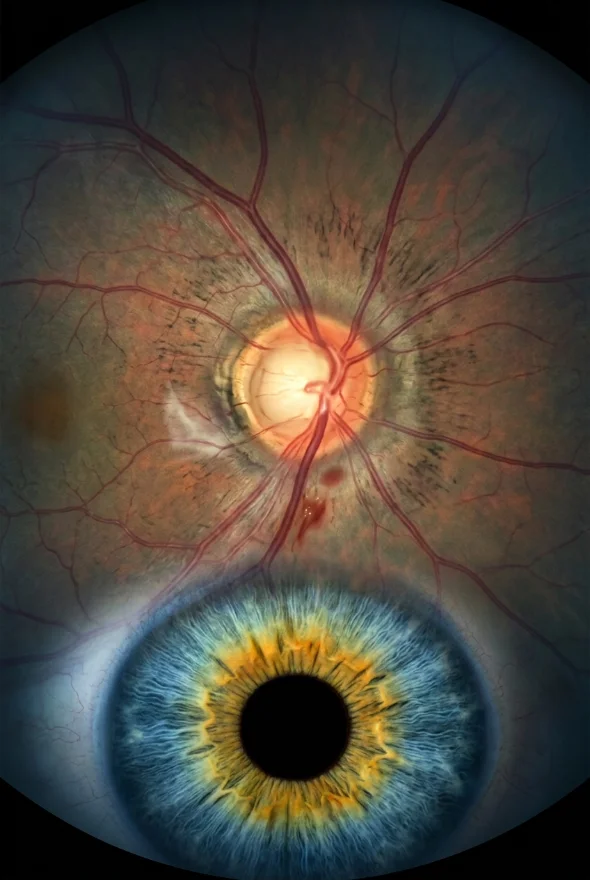
Retina (noun, plural retinas or retinae): the retina is the thin, light-sensitive layer of nerve tissue that lines the inside of the back of the eye. It acts like the sensor of a camera: light-sensing cells called rods and cones turn the image focused by the cornea and lens into electrical signals, and the optic nerve carries those signals to the brain, which turns them into what you see. Retinal diseases such as retinal detachment, macular degeneration and diabetic retinopathy can cause permanent sight loss if they are not found and treated in time.
Retina: key facts
- Where it is
- The inner lining of the back wall of the eye, behind the vitreous gel and in front of the choroid (the blood-rich layer beneath it)
- Thickness
- About 0.5 mm
- Main cells
- Photoreceptors: rods (dim light, side vision) and cones (detail and colour); then bipolar, horizontal and amacrine cells and ganglion cells
- Centre of sharp vision
- The macula, with the fovea at its centre (the fovea is less than a quarter of a millimetre, about 200 µm, across)
- Exit to the brain
- The optic nerve, at the optic disc
- Layers
- Ten, counting the pigment epithelium and the inner limiting membrane
- Blood supply
- Two sources: the central retinal artery feeds the inner retina, and the choroid feeds the photoreceptors (it receives the larger share of the eye's blood flow)
- Specialist
- Retina specialist (ophthalmologist); at Laser Istanbul Clinic, Doç. Dr. Muhammet Derda Özer and Op. Dr. Aysan Bayram

What is the retina, and where is it in the eye?
The retina is the layer of cells lining the back wall inside the eye that senses light and sends signals to the brain so that you can see, as the American Academy of Ophthalmology (AAO) puts it. It is only about 0.5 mm thick, yet it holds the first stages of the whole visual system: light capture, early image processing and the cables (nerve fibres) that leave the eye.
The retina sits at the very back of the eyeball. In front of it is the vitreous, the clear gel that fills the middle of the eye. Behind it is the choroid, a layer of blood vessels that feeds the outer retina, and then the white outer wall of the eye, the sclera. Light enters through the cornea, passes through the pupil and the lens, crosses the vitreous and has to travel through the thickness of the retina before it reaches the rods and cones, which lie at the back against the pigment layer.
Retina, macula and fovea: what is the difference? The retina is the whole lining. The macula is its small central area, responsible for the sharp, straight-ahead vision you use to read, recognise faces and see colour. The fovea is the tiny pit at the very centre of the macula, less than a quarter of a millimetre across, where cones are packed most tightly. Everything outside the macula is the peripheral retina, which gives side vision and helps you see in dim light.
Retina function: how the retina creates vision
The retina does four jobs, in this order.
1. It captures light. The photoreceptors convert light into electrical signals. There are two kinds. Rods work in dim light; they contain the pigment rhodopsin and give you night vision and side vision. Cones work in bright light, give sharp detail and let you see colour; people normally have three types, sensitive mainly to red, green and blue light. Cones dominate the centre of the retina and rods dominate the periphery.
2. It processes the signal. Before anything leaves the eye, bipolar, horizontal and amacrine cells begin sorting the information, for example picking out edges and contrast.
3. It sends the result to the brain. The output cells, the ganglion cells, send their long fibres across the surface of the retina and gather at the optic disc, where they leave the eye as the optic nerve. The optic disc has no light-sensing cells, which is why everyone has a natural blind spot there.
4. It is fed by a dedicated blood supply. The retina is a very energy-hungry tissue. According to the Webvision retina textbook, the choroid receives the greatest share of the eye's blood flow (65–85%) and nourishes the photoreceptors, while the central retinal artery supplies the inner retina with 20–30%. That is why blood-vessel disease, such as diabetes or a blocked retinal vein, shows up in the retina, and why a detached retina, cut off from the choroid beneath it, is an emergency.
The layers of the retina
Under a microscope, and on an OCT scan, the retina shows ten layers. Listed from the back of the eye (outermost) to the front (innermost, facing the vitreous):
| # | Layer | What it contains or does |
|---|---|---|
| 1 | Retinal pigment epithelium (RPE) | A single layer of pigmented cells that supports and nourishes the rods and cones |
| 2 | Photoreceptor layer | The light-sensing outer parts of the rods and cones |
| 3 | Outer (external) limiting membrane | Junctions between Müller support cells and the photoreceptors |
| 4 | Outer nuclear layer | Cell bodies of the rods and cones |
| 5 | Outer plexiform layer | Connections between photoreceptors and bipolar and horizontal cells |
| 6 | Inner nuclear layer | Cell bodies of bipolar, horizontal and amacrine cells |
| 7 | Inner plexiform layer | Connections between bipolar cells and ganglion cells |
| 8 | Ganglion cell layer | Cell bodies of the ganglion cells, the output neurons of the retina |
| 9 | Nerve fibre layer | Ganglion cell fibres running to the optic nerve |
| 10 | Inner limiting membrane (ILM) | A thin membrane facing the vitreous |
Strictly speaking, the RPE is the outer wall on which the nerve retina rests. A retinal detachment is a separation between layers 1 and 2: the nerve retina lifts away from the pigment layer that feeds it. In macular hole surgery, the surgeon often peels the inner limiting membrane around the hole to help it close.
Retinal diseases: common conditions that affect the retina
Retinal tear and retinal detachment
With age, the vitreous gel shrinks and pulls away from the retina (posterior vitreous detachment). This is common and usually harmless, but sometimes the gel sticks and pulls hard enough to tear the retina, letting fluid pass behind it so the retina peels away. According to the AAO, you are at higher risk if you are short-sighted, have had cataract or other eye surgery or a serious eye injury, or have a family history of detachment. EyeWiki (AAO) gives an incidence of about 1 in 10,000 people. Retinal detachment is an emergency: untreated, it can lead to permanent vision loss.
Age-related macular degeneration (AMD)
AMD damages the macula and blurs central vision while side vision is kept. The US National Eye Institute (NEI) says about 11 million people in the United States have it. Dry AMD is the more common form: the macula thins slowly over years, and there is no treatment for late-stage dry AMD. Wet AMD is less common and faster: abnormal blood vessels grow under the macula and damage it, and it can be treated with anti-VEGF injections. Risk factors are age (55 and over), a family history and smoking.
Diabetic retinopathy
High blood sugar damages the tiny blood vessels in the retina. Early stages usually cause no symptoms, which is why screening matters; later the vessels leak or new, fragile ones grow, causing blurred vision, floaters and bleeding inside the eye. The NEI notes that anyone with type 1, type 2 or gestational diabetes is at risk, and that about 1 in 15 people with diabetes will develop diabetic macular oedema (swelling of the macula). The NHS invites people with diabetes aged 12 and over for eye screening every 1–2 years.
Retinal vein occlusion
A blockage in the central retinal vein or one of its branches causes bleeding and swelling in the retina, usually with sudden loss of vision in one eye. According to EyeWiki, high blood pressure, diabetes, glaucoma and cardiovascular disease are the main risk factors.
Macular hole and epiretinal membrane (macular pucker)
A macular hole is a small break in the macula, causing blurred or distorted central vision; an epiretinal membrane is a thin scar-like film on the macula that wrinkles it. Both are usually treated with vitrectomy surgery.
Retinitis pigmentosa and inherited retinal disease
Retinitis pigmentosa is a group of rare inherited conditions in which retinal cells gradually deteriorate. Night vision is usually lost first, often in childhood, followed by a narrowing of side vision ("tunnel vision"). The NEI states there is no cure for retinitis pigmentosa; vision aids, regular monitoring and genetic counselling help.
Other conditions
Central serous chorioretinopathy, cytomegalovirus retinitis, retinopathy of prematurity, retinoblastoma (a rare childhood eye cancer) and Usher syndrome are also listed by the AAO among retinal diseases.
Damaged retina symptoms: warning signs to know
Many retinal diseases cause no pain, and early diabetic retinopathy and early AMD cause no symptoms at all. Watch for these signs:
| Symptom | What it can point to |
|---|---|
| New floaters (dots, lines, cobwebs), especially a sudden shower | Posterior vitreous detachment, retinal tear or detachment, bleeding |
| Flashes of light | Vitreous pulling on the retina; tear or detachment |
| A dark curtain, shadow or grey area spreading across your vision | Retinal detachment: emergency |
| Blurred or distorted central vision; straight lines look wavy | Macular disease: AMD, macular hole, epiretinal membrane, macular oedema |
| Sudden painless loss of vision in one eye | Retinal vein or artery blockage, bleeding, detachment: emergency |
| Trouble seeing at night, narrowing of side vision | Retinitis pigmentosa and other inherited conditions |
| Worsening vision with long-standing diabetes | Diabetic retinopathy or macular oedema |
The NHS and the AAO give the same advice for the first three rows: new flashes, floaters or a shadow need an eye examination straight away.
How is the retina examined?
An eye specialist uses a mix of examinations and scans, most of them quick and painless:
- Dilated retinal examination. Drops widen the pupil so the doctor can see the whole retina, including its far edge, using a bright light and a special lens. The NEI calls it the only way to check for many eye diseases early. Vision is blurry and light-sensitive for a few hours afterwards, so do not plan to drive straight afterwards.
- Retinal photography. A photograph of the back of the eye gives a record to compare over time. NHS diabetic eye screening uses photographs of the back of the eye.
- Optical coherence tomography (OCT). A scan, like an ultrasound that uses light, that shows a cross-section of the retinal layers. It is the key test for macular holes, epiretinal membranes, macular oedema and AMD.
- Ultrasound. If blood or a dense cataract blocks the view, ultrasound can show whether the retina is detached.
- Electrical and genetic tests. The NEI lists electroretinography (ERG) and genetic testing among the tests used for retinitis pigmentosa.
Photographs and OCT are valuable, but they do not replace a dilated examination when a tear or detachment is suspected. At Laser Istanbul Clinic, a retina assessment includes a dilated examination by your surgeon and the relevant scans, and your surgeon can review your existing scans before you travel.
Retina treatment: how retinal conditions are treated
| Treatment | What it does | Typically used for |
|---|---|---|
| Observation and monitoring | Regular examinations and scans | Early diabetic retinopathy, early dry AMD, harmless floaters, small lesions that are not threatening vision |
| Laser (retinal photocoagulation) or freezing (cryotherapy) | Seals the retina around a tear, or treats leaking and abnormal vessels | Retinal tears, diabetic retinopathy |
| Anti-VEGF injections | Medicine injected into the eye to stop abnormal vessel growth and reduce macular swelling | Wet AMD, diabetic macular oedema, retinal vein occlusion |
| Steroid implants or injections | Reduce swelling and inflammation | Macular oedema not responding to anti-VEGF |
| Pneumatic retinopexy | A gas bubble pushes the retina back into place | Selected retinal detachments |
| Scleral buckle | A band sewn on the outside of the eye pushes the wall inwards | Retinal detachment |
| Vitrectomy | The vitreous gel is removed so the surgeon can repair the retina directly; the eye is filled with gas, air or silicone oil | Retinal detachment, macular hole, macular pucker, bleeding inside the eye, advanced diabetic disease |
Retinal detachment surgery. EyeWiki (AAO) reports that anatomic success exceeds 90% with modern surgical techniques, and the AAO notes that about 1 in 20 patients need a second operation. Sight begins to improve about four to six weeks after surgery, but the retina can keep healing for a year or more. If gas is used in the eye, you must not fly or go to high altitude until it has gone.
Can a damaged retina repair itself? Not in humans. Fish such as zebrafish can regrow retinal nerve cells, but the human retina cannot, and the AAO states there are no FDA-approved stem cell treatments for eye disease. Treatment aims to stop damage getting worse and to repair what can be repaired. This is also why early detection matters so much.
Treatment with us: retina care at Laser Istanbul Clinic is led by Doç. Dr. Muhammet Derda Özer (Doç. = Associate Professor), who treats retinal disease, uveitis and ocular tumours, together with Op. Dr. Aysan Bayram, who also treats retinal disease and speaks English. If you already have a retina diagnosis and want a second opinion or planned surgery, you can send your scans before you travel.

When to see a doctor about your retina
Get seen the same day, at the nearest eye emergency service, if you have:
- sudden flashes of light, or a sudden shower of new floaters
- a dark shadow or "curtain" moving across your vision
- a sudden drop or loss of vision in one eye
- new blurring with eye pain, or any of these symptoms after eye surgery or an eye injury
Do not wait to travel for treatment. If you are abroad and think you may have a retinal tear or detachment, go to a local emergency eye service first.
Book a routine appointment if you notice:
- straight lines that look wavy or a blurred patch in the middle of your vision
- gradual loss of side vision or difficulty seeing at night
- a long history of diabetes with no recent eye screening, or any change in your vision
- a family history of retinal detachment or inherited retinal disease
If you are short-sighted, have diabetes, are over 55 or have had eye surgery, regular eye checks that include a look at the retina are especially worthwhile.
Frequently asked questions
The retina is the thin, light-sensitive layer of tissue lining the back of the eye. It converts light into electrical signals that travel through the optic nerve to the brain.
It captures the image formed by the cornea and lens, uses rods and cones to turn light into signals, does the first stage of processing, and sends the result to the brain. Cones give sharp central and colour vision; rods give night and side vision.
There are ten, from the back of the eye forwards: retinal pigment epithelium, photoreceptor layer, outer limiting membrane, outer nuclear layer, outer plexiform layer, inner nuclear layer, inner plexiform layer, ganglion cell layer, nerve fibre layer and inner limiting membrane.
The retina is the whole light-sensitive lining of the back of the eye. The macula is its small central area that gives sharp, detailed vision, and the fovea is the tiny pit at its centre.
New floaters, flashes of light, a dark curtain across your vision, blurred or wavy central vision and sudden loss of vision can all point to retinal problems. Some conditions, including early diabetic retinopathy and early AMD, cause no symptoms, so routine examination matters.
No, not in humans. The retina cannot regrow lost nerve cells, so treatment aims to prevent further damage and repair what can be repaired. A detached retina in particular needs surgery, and the sooner it is treated the better the chance of saving vision.
By surgery: a gas bubble (pneumatic retinopexy), a scleral buckle or vitrectomy, with laser or freezing sometimes used to seal the retina in place. The choice depends on where the tear is and the type of detachment.
No. The drops may sting briefly. Your vision will be blurry and light-sensitive for a few hours, so avoid driving until it clears.
Partly. Controlling blood sugar, blood pressure and cholesterol and not smoking lowers the risk of diabetic retinopathy, and attending screening lets treatment start early. Retinal tears and detachments generally cannot be prevented, so knowing the warning signs is the best protection.
Sources for the facts on this page
- American Academy of Ophthalmology. What Is the Retina? https://www.aao.org/eye-health/anatomy/retina-list
- Kolb H. Simple Anatomy of the Retina. Webvision, University of Pittsburgh. https://www.webvision.pitt.edu/book/part-i-foundations/simple-anatomy-of-the-retina/
- Webvision. Photoreceptors (rods and cones). https://www.webvision.pitt.edu/book/part-ii-anatomy-and-physiology-of-the-retina/photoreceptors/
- National Eye Institute (NIH). How the eyes work. https://www.nei.nih.gov/learn-about-eye-health/healthy-vision/how-eyes-work
- American Academy of Ophthalmology. Detached or torn retina. https://www.aao.org/eye-health/diseases/detached-torn-retina
- NHS. Detached retina (retinal detachment). https://www.nhs.uk/conditions/detached-retina-retinal-detachment/
- NHS. Floaters and flashes in the eyes. https://www.nhs.uk/conditions/floaters-and-flashes-in-the-eyes/
- EyeWiki (AAO). Retinal Detachment. https://eyewiki.org/Retinal_Detachment
- National Eye Institute (NIH). Age-related macular degeneration. https://www.nei.nih.gov/learn-about-eye-health/eye-conditions-and-diseases/age-related-macular-degeneration
- National Eye Institute (NIH). Diabetic retinopathy. https://www.nei.nih.gov/learn-about-eye-health/eye-conditions-and-diseases/diabetic-retinopathy
- NHS. Diabetic retinopathy. https://www.nhs.uk/conditions/diabetic-retinopathy/
- EyeWiki (AAO). Retinal Vein Occlusion. https://eyewiki.org/Retinal_Vein_Occlusion
- National Eye Institute (NIH). Retinitis pigmentosa. https://www.nei.nih.gov/learn-about-eye-health/eye-conditions-and-diseases/retinitis-pigmentosa
- National Eye Institute (NIH). Get a dilated eye exam. https://www.nei.nih.gov/learn-about-eye-health/healthy-vision/get-dilated-eye-exam
- EyeWiki (AAO). Optical Coherence Tomography (retinal layers on OCT). https://eyewiki.aao.org/Optical_Coherence_Tomography
- American Academy of Ophthalmology. Stem Cell Therapy for Eye Disease: What You Need to Know (4 Jun 2019). https://www.aao.org/eye-health/tips-prevention/stem-cell-therapy-eye-disease
- Campbell LJ, Levendusky JL, Steines SA, Hyde DR. Retinal regeneration requires dynamic Notch signaling. Neural Regen Res 2022 (PMC8643038): zebrafish Müller glia regenerate retinal neurons. https://pmc.ncbi.nlm.nih.gov/articles/PMC8643038/
Worried about your retina, or told you need retina surgery?
Send us your scans or eye report. Doç. Dr. Muhammet Derda Özer or Op. Dr. Aysan Bayram will review them and tell you which treatments may suit your eyes, with no obligation.
3 eye surgeons
Your case is reviewed by a named surgeon, not a salesperson. Reply within {{response time}}.
 Prof. Dr. Ahmet Satıcı
 Doç. Dr. Muhammet Derda Özer
 Op. Dr. Aysan Bayram