
- Home
- Knowledge centre
- Eye glossary A–Z
- Keratoconus: What It Is, Symptoms, Causes and Treatment
Keratoconus: What It Is, Symptoms, Causes and Treatment
Medically reviewed by {{surgeon}}, {{surgeon's title and specialty}}, on {{review date}}. Last updated {{date}}.
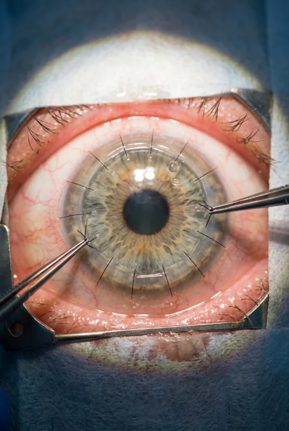
Keratoconus (noun, pronounced ker-uh-toh-KOH-nus): keratoconus is an eye condition in which the clear front surface of the eye, the cornea, gradually thins and bulges forward into a cone shape. The cone changes how light focuses inside the eye, causing blurred and distorted vision that glasses correct less and less well. It usually starts in the teens or early twenties and is managed by a cornea specialist; at Laser Istanbul Clinic, keratoconus care is led by Doç. Dr. Muhammet Derda Özer (Doç. means Associate Professor).
Keratoconus: key facts
- What happens
- The cornea thins and steepens into a cone, causing irregular astigmatism and short-sightedness that keep changing
- Usual age at start
- Late teens to early twenties; it can progress over 10–20 years (AAO)
- How common
- Often quoted as about 1 in 700 people; published estimates range from 0.17 to 40 per 1,000 depending on how it is measured and where (EyeWiki; 2026 clinical practice guideline)
- Main known risk factors
- Eye rubbing, eye allergy, family history, some connective-tissue conditions, Down syndrome
- Key test
- Corneal topography or tomography (a map of the cornea's shape and thickness)
- Main treatments
- Glasses and special contact lenses; corneal cross-linking to stop progression; ring segments; corneal transplant in advanced cases
- Specialist
- Cornea specialist (ophthalmologist); at Laser Istanbul Clinic, Doç. Dr. Muhammet Derda Özer
What is keratoconus?
Keratoconus is a progressive disease of the cornea, the dome-shaped clear window at the front of the eye. The cornea normally holds its round shape because its collagen fibres are strong and evenly arranged. In keratoconus those fibres weaken, so the cornea slowly thins, usually in the lower centre, and pushes forward into a cone. Because the cornea provides most of the eye's focusing power, even a small change in its shape matters: light no longer meets in a single point, and vision becomes blurred, doubled or streaked.
The word comes from the Greek kerato (cornea) and konos (cone). It is not contagious, and it is not caused by wearing glasses or contact lenses.
How common is keratoconus?
EyeWiki, the American Academy of Ophthalmology's online reference, says the prevalence is often reported as 1 in 700, with higher rates in Middle Eastern, Indian, Chinese and Australian populations. A 2026 clinical practice guideline in Clinical Ophthalmology notes that published estimates range from 0.17 to 40 cases per 1,000 people, because studies use different diagnostic criteria and imaging technology. Better scanners now find milder cases that were missed in the past.
Does keratoconus get worse over time?
Often, yes, at least for a while. The American Academy of Ophthalmology (AAO) says keratoconus typically begins in the late teens to early twenties and can progress over 10 to 20 years. EyeWiki describes onset in early adolescence with progression into the mid-twenties and thirties, although it can start earlier or continue later. It tends to progress faster in children and young people: a 2026 European consensus statement on paediatric keratoconus cites progression in 88% of children in published studies, with some eyes steepening by up to 5 dioptres in a year. How an individual eye will behave cannot be predicted without regular scans.
What causes keratoconus?
The exact cause is not known. Current understanding is that keratoconus results from a combination of inherited tendencies and things that physically or chemically stress the cornea.
| Factor | What the evidence says |
|---|---|
| Eye rubbing | The strongest risk factor you can change. The 2026 guideline reports an odds ratio of 5.22 for eye rubbing; the paediatric consensus says children who rub their eyes are about three times as likely to develop keratoconus. The AAO advises avoiding eye rubbing because it can damage thin corneal tissue and make symptoms worse |
| Family history | About 1 in 10 people with keratoconus have an affected parent (AAO). The 2026 guideline reports an odds ratio of 6.67 for family history |
| Eye allergy and atopy | Allergic eye disease raises the risk (odds ratio 2.21 in the 2026 guideline), mainly because it makes people rub their eyes. Controlling the allergy with drops is part of treatment |
| Connective-tissue conditions | Marfan syndrome and Ehlers-Danlos syndrome are associated with keratoconus (AAO) |
| Down syndrome | Listed by EyeWiki as a risk factor |
| Other associations | EyeWiki also lists sleep apnoea, hypothyroidism and hormonal changes during pregnancy. These are associations, not proven causes |
Not a cause: diabetes and sex showed no significant association in the 2026 guideline's review.

What are the symptoms of keratoconus?
Early keratoconus can cause no symptoms or just a little blur, which is why it is often found at a routine eye test or before laser eye surgery. As the cone grows, the AAO lists these symptoms:
- Early: mild blurring, slight distortion (straight lines look bent or wavy), more sensitivity to light and glare, and sometimes eye redness or swelling.
- Later: more severe blurring and distortion, increasing short-sightedness and astigmatism that need frequent changes of glasses, and difficulty wearing contact lenses because they fit poorly or feel uncomfortable.
EyeWiki sums the picture up as progressive changes in vision that are not easily corrected with glasses, with a prescription that will not stay stable.
Acute hydrops: a sudden worsening
Occasionally a break forms in Descemet's membrane, the thin inner layer of the cornea, letting fluid from inside the eye enter the corneal tissue. EyeWiki calls this acute hydrops: the cornea thickens and clouds, and vision drops with light sensitivity, watering and pain. It is managed with medication and sometimes a pressure patch, and patients are told to avoid vigorous rubbing while it settles. Sudden clouding of vision in a person with keratoconus needs same-day eye care.
How is keratoconus diagnosed?
Keratoconus is diagnosed by an eye specialist during an eye examination. The doctor examines the cornea and measures its curvature, and the AAO notes that a special computer is used to map the cornea's surface. The tests are quick and painless:
| Test | What it shows |
|---|---|
| Visual acuity and refraction | How well you see with glasses; irregular astigmatism that will not correct fully is a clue |
| Retinoscopy | A "scissoring" reflex of light, a sign of an irregular cornea (EyeWiki) |
| Slit-lamp examination | A microscope view of the cornea. Specialists look for Vogt's striae (fine, roughly vertical lines in the corneal tissue), a Fleischer ring (an iron deposit around the base of the cone) and Munson's sign (the lower eyelid bulging out when you look down) |
| Corneal topography and tomography | Maps of the front surface (topography) and of the front and back surfaces with thickness (tomography). They detect early and "forme fruste" (subclinical) keratoconus; maximum keratometry (Kmax) is one of the main readings used to track progression |
| Pachymetry | Measures corneal thickness, usually thinnest at the cone |
| Epithelial thickness mapping | Maps the corneal surface layer; EyeWiki lists it as complementary data for early disease |
Why scans matter: because topography can pick up very early keratoconus, it is essential before any laser eye surgery. The AAO lists keratoconus among the reasons a person is not a candidate for LASIK.
How often is keratoconus checked?
A 2026 clinical practice guideline recommends risk-based follow-up: about every 12 months for mild, stable adult disease, every 6 months when allergy or eye rubbing is present, and every 3–4 months for young patients, documented progression or ectasia after refractive surgery. A European consensus for children advises checks every 3 months at ages 3–12, every 4 months at ages 12–20, every 6 months at ages 20–40 and yearly after 40, extending the interval when the cornea is stable.
Should children be screened?
The paediatric consensus advises corneal imaging for children aged 6 to 18 years whose refraction shows astigmatism above 2 dioptres, and examination around the ages of 8–13 when there is a family history. Screening below age 12 is generally discouraged because of the risk of false positives. Parents of a young person whose prescription keeps changing should ask for a scan.
How is keratoconus treated?
Treatment has two aims: to stop the cornea getting worse (cross-linking) and to give the clearest vision possible (glasses, lenses, rings or transplant). The right plan depends on the stage and on whether the disease is progressing.
| Treatment | What it does | Typically used for |
|---|---|---|
| Glasses | Correct short-sightedness and regular astigmatism | Early keratoconus |
| Soft, rigid gas-permeable (RGP), hybrid and scleral contact lenses | A rigid or scleral lens forms a smooth new optical surface over the irregular cornea. EyeWiki describes the usual progression from soft toric lenses to RGP and then specialty designs such as semi-scleral, scleral, hybrid and piggyback lenses | Most patients, throughout the disease |
| Corneal cross-linking (CXL) | Riboflavin (vitamin B2) drops plus ultraviolet-A light strengthen the corneal tissue to halt further bulging | Progressive keratoconus |
| Intrastromal corneal ring segments (Intacs) | Small curved implants placed in the cornea to flatten the cone and regularise its shape | Mild to moderate keratoconus, often when contact lenses are not tolerated |
| Corneal transplant (keratoplasty) | Replaces diseased corneal tissue with healthy donor tissue | Advanced keratoconus or scarring when other measures are not enough |
What is corneal cross-linking?
Corneal cross-linking strengthens the cornea by creating new bonds between its collagen fibres. The surgeon applies riboflavin drops to the cornea and exposes it to ultraviolet-A light. EyeWiki states that the goal is to halt changes in corneal shape if the disease is progressing, not to guarantee better vision, so cross-linking is most valuable when done before the cornea has become badly distorted. It does not remove the cone, but it is the treatment designed to slow or stop the disease itself.
- Epithelium-off ("epi-off") removes the thin surface layer first so the riboflavin can soak in. The 2026 guideline reports it gives greater reduction in maximum keratometry but carries higher complication risks.
- Epithelium-on (transepithelial) leaves the surface in place. The guideline says it has a better safety profile but a less pronounced effect.
- Accelerated protocols shorten the ultraviolet exposure. The paediatric consensus calls an accelerated epi-off protocol of 9 mW for 20 minutes (5.4 J/cm²) the gold standard in children, and recommends treating at diagnosis even before progression is shown, because young eyes progress so often. The 2026 guideline notes that conventional (slower) cross-linking may be preferred when a greater flattening effect is wanted.
- Regulation: EyeWiki records that the US Food and Drug Administration approved Photrexa/KXL cross-linking in 2016 and an epithelium-on system, Epioxa HD, in October 2025.
What are corneal ring segments?
Intacs and similar segments are placed in the cornea to flatten the cone. EyeWiki says they are approved for mild-to-moderate keratoconus in people who cannot tolerate contact lenses and need enough corneal thickness (above 450 micrometres). The 2026 guideline lists common adverse events as white deposits (5.75%), surface defects (5.65%), segment extrusion (2.11%) and decentration (1.41%). Ring segments can be combined with cross-linking when both strengthening and reshaping are wanted.
When is a corneal transplant needed?
A transplant is reserved for advanced disease, deep scarring or when lenses no longer work. Two main operations are used:
| Operation | What is replaced | Notes |
|---|---|---|
| DALK (deep anterior lamellar keratoplasty) | The front and middle layers; your own inner layer (endothelium) stays | Preferred in keratoconus when the endothelium is healthy. The 2026 guideline reports lower rates of raised eye pressure, cataract and graft rejection than full-thickness grafts |
| PK (penetrating keratoplasty) | The full thickness of the cornea | Still used where DALK is not possible; EyeWiki notes it has high success rates |
Rejection can happen after any transplant, so a red, painful, light-sensitive or blurred eye after a transplant needs urgent attention.
Treatment with us: cross-linking, ring segments and corneal transplants for keratoconus are led by Doç. Dr. Muhammet Derda Özer, who trained at Istanbul University's Faculty of Medicine, holds the International Council of Ophthalmology's FICO qualification, and whose published research includes corneal cross-linking for keratoconus in young people. Surgery takes place in a fully equipped, Ministry of Health–licensed operating theatre in Istanbul.
Can I have laser eye surgery with keratoconus?
No. Laser vision correction removes corneal tissue, which would further weaken an already thin and unstable cornea. The AAO lists keratoconus among the reasons a person is not a candidate for LASIK. Keratoconus is treated with the approaches above instead.
Can keratoconus be cured, and does it cause blindness?
Keratoconus cannot be reversed to a normal cornea, but progression can often be halted and vision improved with the treatments above. Early diagnosis and cross-linking before the cone becomes severe offer the best chance of avoiding a transplant. Left unmonitored and untreated, it can cause lasting loss of clear vision.
What can I do myself?
- Don't rub your eyes. Rubbing is the strongest modifiable risk factor and the AAO says it can damage thin corneal tissue. If itching is the trigger, ask about allergy drops; the 2026 guideline lists antihistamine and mast-cell-stabilising drops as first-line.
- Keep scheduled scans. Progression is found by comparing topography over time, not by how you feel.
- Have your lenses checked. A badly fitting lens is a common reason keratoconus patients become uncomfortable.
- Report any sudden change. A sudden cloudy cornea, new pain or a red, light-sensitive eye needs same-day care.
- Tell any eye surgeon you have keratoconus before any procedure, and have scans done before considering laser surgery.

When to see a doctor about keratoconus
Seek same-day care from an eye doctor or eye emergency service if you have:
- sudden clouding or a sharp drop in vision in an eye known to have keratoconus (possible acute hydrops)
- a painful red eye with sensitivity to light, especially in a contact-lens wearer (take your lenses out and don't put them back in)
- redness, pain, light sensitivity or blurred vision after a corneal transplant
Book an appointment soon if you notice:
- blurred or distorted vision that keeps getting worse, or a glasses prescription (especially astigmatism) that changes every year, particularly in a teenager or young adult
- ghosting or streaking around lights that glasses do not fix
- contact lenses that suddenly stop fitting or feel uncomfortable
- a family member with keratoconus and any of the symptoms above
If you already have a diagnosis and want a second opinion on cross-linking, ring segments or a transplant, you can send your scans for review before you travel.
Frequently asked questions
Keratoconus is a condition in which the cornea, the clear front window of the eye, becomes thin and bulges into a cone shape. This distorts how light enters the eye and causes blurred, warped vision that glasses cannot fully correct. It usually begins in the teens or early twenties.
The exact cause is unknown. Eye rubbing, eye allergies and a family history are the best-established risk factors, and about 1 in 10 people with keratoconus have an affected parent. Connective-tissue conditions such as Marfan and Ehlers-Danlos syndromes and Down syndrome are also linked to it.
Mild blurring, slight distortion where straight lines look wavy, more glare and light sensitivity, and a glasses prescription that keeps changing. Early keratoconus may cause no symptoms and can be found only on a corneal scan.
Often, yes. Corneal cross-linking strengthens the cornea and is designed to halt progression, ideally before the cone is severe. It is not guaranteed to improve vision, and it works best when the disease is caught early.
Sometimes. About 1 in 10 people with keratoconus have a parent with it, and the 2026 guideline reports an odds ratio of 6.67 for family history. Relatives, especially teenagers, can ask for a corneal scan.
Yes. It can start in early adolescence or sometimes earlier, and it tends to progress faster in children, so regular scans every few months and early cross-linking are recommended for progressive disease. Children whose prescription keeps changing, especially with rising astigmatism, should be checked.
Often yes, but not ordinary soft lenses once the cone is irregular. Rigid gas-permeable, hybrid and scleral lenses give a smooth surface over the cone and are the main way many patients see clearly.
No. The AAO lists keratoconus among the reasons a person cannot have LASIK. Cross-linking and other keratoconus treatments are used instead.
No. A transplant is for advanced disease, scarring or when lenses no longer work. Early cross-linking is aimed at stopping the disease before that stage.
Total blindness is not the usual outcome, but without monitoring and treatment keratoconus can cause lasting loss of clear vision. With glasses, special contact lenses, cross-linking, ring segments or a corneal transplant, most patients keep useful vision.
Sources for the facts on this page
- American Academy of Ophthalmology. What Is Keratoconus? https://www.aao.org/eye-health/diseases/what-is-keratoconus
- American Academy of Ophthalmology. Keratoconus Diagnosis and Treatment. https://www.aao.org/eye-health/diseases/keratoconus-diagnosis
- EyeWiki (AAO). Keratoconus. https://eyewiki.org/Keratoconus
- Evidence-Based Clinical Practice Guideline for Diagnosis, Treatment, and Follow-Up of Keratoconus Patients. Clinical Ophthalmology 2026. PubMed PMID 42763659; open access: https://pmc.ncbi.nlm.nih.gov/articles/PMC13589533/
- Paediatric Keratoconus: An ERN-EYE Clinical Consensus Statement on Diagnosis, Treatment and Follow-Up Care. Ophthalmology and Therapy 2026. PubMed PMID 42538519; open access: https://pmc.ncbi.nlm.nih.gov/articles/PMC13518778/
- American Academy of Ophthalmology. LASIK — Laser Eye Surgery (candidacy). https://www.aao.org/eye-health/treatments/lasik
Told you have keratoconus, or worried your prescription keeps changing?
Send us your scans or eye report. Doç. Dr. Muhammet Derda Özer, our cornea and keratoconus specialist, will review them and tell you which treatments may suit your eyes, with no obligation.
3 eye surgeons
Your case is reviewed by a named surgeon, not a salesperson. Reply within {{response time}}.
 Prof. Dr. Ahmet Satıcı
 Doç. Dr. Muhammet Derda Özer
 Op. Dr. Aysan Bayram