
- Home
- Knowledge centre
- Eye glossary A–Z
- Eye Lens (Crystalline Lens): Structure, Function and Problems
Eye Lens (Crystalline Lens): Structure, Function and Problems
Medically reviewed by {{surgeon}}, {{surgeon's title and specialty}}, on {{review date}}. Last updated {{date}}.
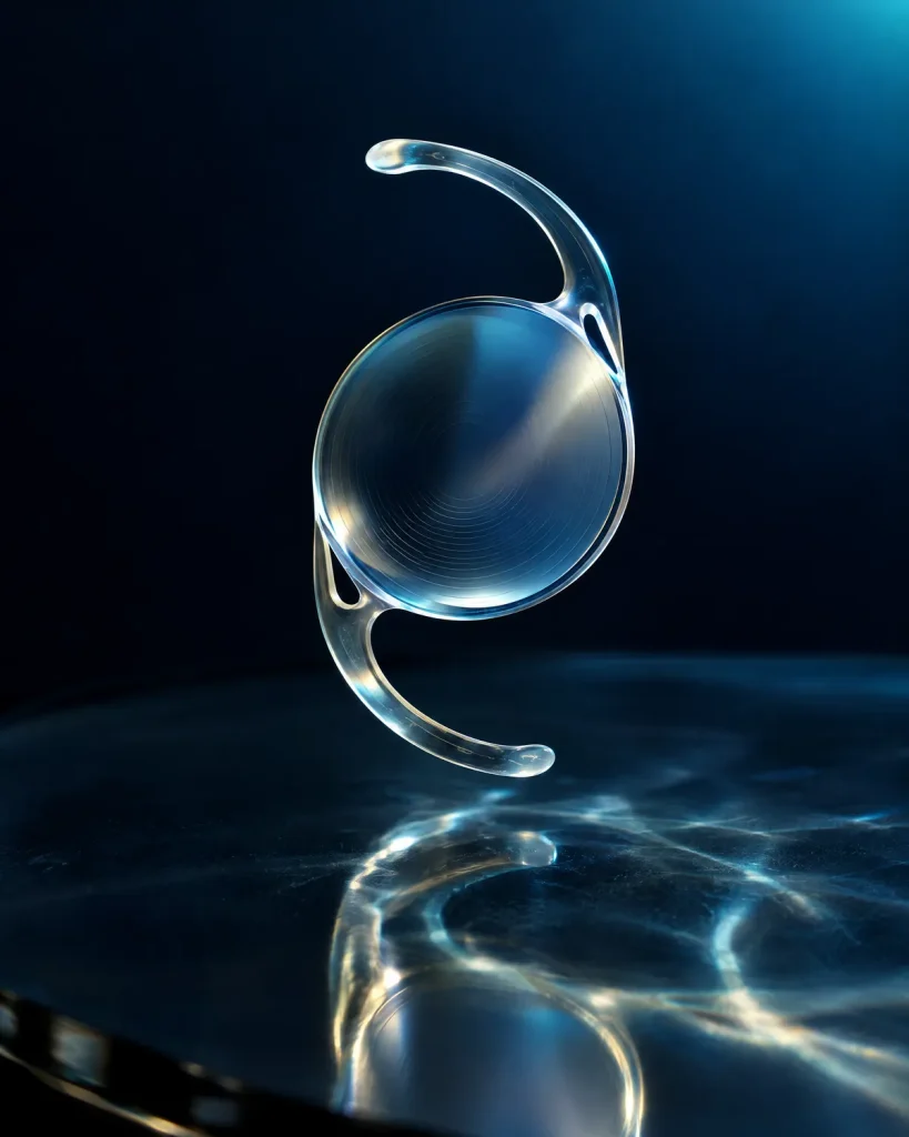
Crystalline lens (noun; also "the lens of the eye" or simply "the eye lens", plural lenses): the clear, flexible, disc-shaped structure that sits directly behind the coloured iris and the pupil. It works with the cornea to focus light on the retina, and it changes shape so that you can switch between seeing far and near. With age the lens stiffens (presbyopia, the reason most people need reading glasses) and can turn cloudy (a cataract), and cataract surgery replaces it with an artificial lens.
In short: the eye lens is the part of the eye that fine-tunes and adjusts focus. If it clouds, the treatment is cataract surgery, which at Laser Istanbul Clinic is performed in Istanbul by Prof. Dr. Ahmet Satıcı (premium and trifocal lenses), Op. Dr. Aysan Bayram or Doç. Dr. (Associate Professor) Muhammet Derda Özer.
Eye lens: key facts
- Where it is
- Behind the iris and pupil, held in place by a ring of fine fibres called zonules that connect it to the ciliary body
- Size
- In a 2021 study of 293 eyes having lens surgery, thickness ranged from 3.6 to 5.7 mm and diameter from 7.5 to 11.9 mm, and both increased with age
- Job
- Fine-tunes the eye's focus and changes shape to focus on near objects; the cornea provides roughly two-thirds of the eye's 60 dioptres of focusing power
- Made of
- A thin outer capsule, a single layer of epithelial cells, and long clear lens fibres packed with crystallin proteins
- Typical age changes
- Stiffens from the 40s (presbyopia); clouds with age (cataract), most often after 60
- Treatment
- Glasses for presbyopia; cataract surgery (or refractive lens exchange) swaps the lens for an artificial intraocular lens (IOL)
- Specialist
- Ophthalmologist; at Laser Istanbul Clinic, Prof. Dr. Ahmet Satıcı, Op. Dr. Aysan Bayram and Doç. Dr. Muhammet Derda Özer all treat cataract
What is the lens of the eye?
The lens of the eye is a transparent, slightly rounded disc a few millimetres thick. It hangs behind the iris. The American Academy of Ophthalmology (AAO) describes it simply as the clear part of the eye behind the coloured iris that helps focus light on the retina so that you can see.
Doctors call it the crystalline lens to separate it from the other "lenses" people talk about: the cornea (the clear front window of the eye, which is also a lens in the optical sense), and the glasses, contact lenses and intraocular lenses (IOLs) that support or replace the natural lens. If you search "eye lens", you will find all of these mixed together. This page covers the natural lens first, then the artificial lenses that replace it.
Where it sits. The lens is held in the centre of the eye by zonules, thin collagen fibres that run from the lens to the ciliary body (the ring of muscle behind the iris). Because the zonules attach to the muscle, the muscle can change the curve of the lens. In front of the lens are the iris and pupil; behind it is the jelly-like vitreous body that fills most of the eye.
What is the eye lens made of? Structure and layers
The lens has three structural parts, as EyeWiki (the AAO's clinical wiki) describes them:
| Part | What it is | Why it matters |
|---|---|---|
| Capsule | A thin, elastic bag around the whole lens; EyeWiki calls it the thickest basement membrane in the body, made mostly of type IV collagen | Holds the lens together and is where the zonules attach. In cataract surgery the capsule is kept, and the new IOL is placed inside it |
| Epithelium | A single layer of cells just under the front of the capsule | New lens fibres grow from these cells |
| Lens fibres | Long, hexagonal cells that lose their nuclei as they mature and are filled with crystallin proteins | The crystallins raise the lens's refractive index, which lets it bend light. The fibres at the centre form the nucleus; the outer fibres form the cortex |
The lens has to stay transparent to work. When its proteins break down and clump, it clouds; this is the mechanism of age-related cataract, according to the AAO and the National Eye Institute (NEI).

What does the eye lens do? Function and accommodation
The lens has two jobs.
1. Focusing light. The cornea provides roughly two-thirds of the eye's total 60 dioptres of focusing power, according to the University of Iowa's eye-care tutorial; the lens supplies the rest and fine-tunes the focus. The National Eye Institute puts it simply: the lens works together with the cornea to focus light correctly on the retina.
2. Changing focus (accommodation). The lens is the part of the eye that adjusts. According to EyeWiki, when you look at something close, the ciliary muscle contracts and moves forward and inward, which reduces tension on the zonular fibres and lets the elastic lens become more curved, which increases its focusing power. When you look into the distance, the muscle relaxes, the zonules tighten and the lens flattens again. This happens automatically, many times a day.
How the lens ages: from flexible to stiff
A young lens is soft and springy. EyeWiki gives the typical range of accommodation as 7–10 dioptres in the first two decades of life, falling to about 0.5 dioptres by age 50. This is presbyopia: the AAO describes it as a normal part of ageing in which the lens becomes more rigid and cannot change shape as easily, usually starting shortly after age 40. EyeWiki puts typical first symptoms at 42–44 years, and complete loss of accommodation at 50–55.
What problems affect the eye lens?
Presbyopia (reading-glasses age)
Presbyopia is the loss of near focus described above. People hold reading material further away and may get headaches or eye strain if it is not corrected. The World Health Organization (WHO) estimates that 826 million people are affected by presbyopia, the main cause of near vision impairment worldwide. Options include reading glasses, varifocals, multifocal contact lenses and, for some people, replacing the lens itself (see the section on artificial lenses below).
Cataract (a cloudy lens)
A cataract is a clouding of the natural lens. The NHS describes it as the lens becoming cloudy, causing blurred vision and loss of sight. The NEI says that in age-related cataract, proteins in the lens break down and clump, starting around age 40, and clouding progresses slowly over time; by age 80, more than half of Americans either have a cataract or have had surgery for one. The WHO estimates that 94 million people worldwide have distance vision impairment or blindness from cataract.
EyeWiki lists three common age-related types, which affect different parts of the lens:
| Type | Where it forms | Typical effect |
|---|---|---|
| Nuclear sclerotic | The centre (nucleus) hardens and yellows | Progressive short-sightedness, poorer night vision, duller colours |
| Cortical | Spoke-like opacities in the outer cortex | Glare, with halos or streaks around lights |
| Posterior subcapsular | Under the back of the capsule | Progresses faster; particularly affects near vision |
Other forms include traumatic cataract (after an injury or radiation), congenital cataract (present at birth) and cataract linked to diabetes. Risk factors listed by the NHS, AAO and NEI include age over 60, family history, diabetes, smoking, long-term steroid use, eye injury or earlier eye surgery, and sun exposure without eye protection. EyeWiki states that no medical treatment prevents cataract from developing; surgery is the definitive treatment.
Lens dislocation (ectopia lentis)
If the zonules are weak or damaged, the lens can shift out of position (subluxation) or move completely. According to EyeWiki, injury is the most common cause, and the most common inherited cause is Marfan syndrome (about 75% of people with it); homocystinuria is the second most common (about 90% of those affected). The main symptom is reduced vision, including double vision in one eye. Treatment depends on the cause and degree: glasses or contact lenses, or surgery to remove the lens and use a supported artificial lens.
Cloudy capsule after cataract surgery (posterior capsule opacification, PCO)
After cataract surgery the capsule is left in place to hold the new lens. EyeWiki says that leftover lens cells can grow across the back of the capsule and cloud it, in 20–50% of patients within 2 to 5 years. The NHS notes this can appear months or years after the operation. It is treated with a quick laser procedure (Nd:YAG capsulotomy), which makes a small opening in the cloudy capsule.
Lens problem symptoms: when your eye lens is the cause
| Symptom | What it can point to |
|---|---|
| Need to hold things further away to read; eye strain up close, from the early or mid-40s | Presbyopia |
| Blurred, hazy or "foggy windshield" vision that glasses no longer fix | Cataract |
| Glare from headlights, halos around lights, poor night vision | Cataract, or a side effect of some lens implants |
| Colours look faded or yellowish | Nuclear cataract |
| Double or ghosted images in one eye | Cataract; dislocated lens |
| Frequent changes to your glasses prescription | Cataract (especially growing short-sightedness) |
| Blurring again months or years after cataract surgery | Posterior capsule opacification |
Cataracts develop slowly, and the NEI notes that early cataracts produce no symptoms, so many people notice a change only when it is well established.
How is the eye lens examined?
Lens problems are usually diagnosed with a quick eye examination:
- Visual acuity and refraction. Reading the chart and checking whether new glasses fix your vision.
- Slit-lamp examination. A microscope with a bright beam of light that lets the doctor look at the lens in cross-section and see any clouding and its type.
- Dilated examination. Drops widen the pupil so that the whole lens and the retina behind it can be checked, which also helps rule out other causes of blurred vision.
- Biometry (for surgery). Before cataract or lens replacement surgery, the length of the eye and the curve of the cornea are measured so that the right power of artificial lens can be chosen. At Laser Istanbul Clinic, corneal topography, OCT scanning and lens-power biometry are part of every pre-operative examination in Istanbul, and your surgeon reviews your existing scans before you book.
How is the eye lens treated and replaced?
Presbyopia is corrected with reading glasses, varifocals, monovision or multifocal contact lenses, with prescription eye drops that shrink the pupil and allow close-up focus for up to six hours a day, or with surgery (reshaping the cornea, or refractive lens exchange with multifocal implants), according to the AAO.
Early cataract can be managed with new glasses and better lighting. The AAO advises surgery when a cataract keeps you from doing things you want or need to do; the NHS says cataracts do not usually need treatment straight away.
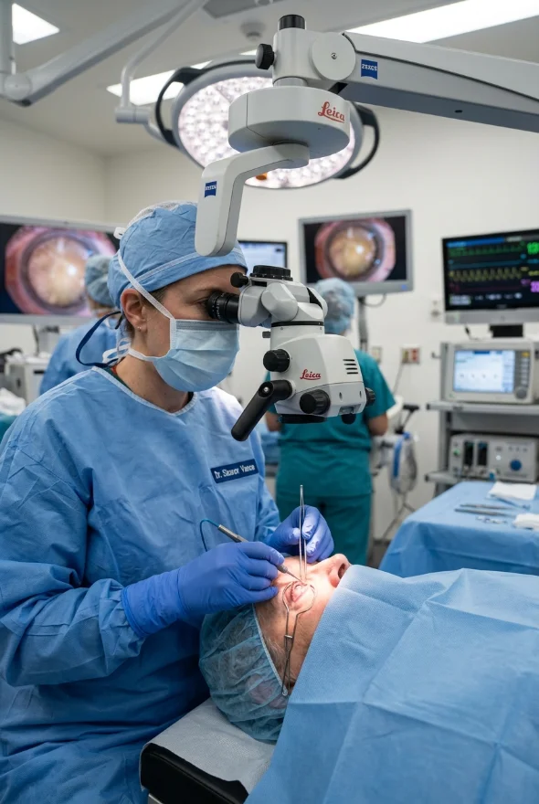
Cataract surgery removes the cloudy lens and replaces it with an artificial intraocular lens (IOL). In the standard technique (phacoemulsification), the surgeon makes a tiny cut, may use ultrasound to break up the lens, removes it and places the IOL inside the capsule. The NHS says the operation takes between 20 and 45 minutes, is done under local anaesthetic with same-day discharge, and eye drops are used for about 4 weeks. EyeWiki reports an overall success rate of 97% or higher; the NHS says serious complications are rare, and lists vision problems, retinal detachment, infection and posterior capsule opacification as possible risks.
Refractive lens exchange (RLE) is the same operation done before a cataract has formed, to reduce dependence on glasses. The Royal College of Ophthalmologists states that RLE uses the same procedure as cataract surgery.
Types of artificial eye lens (IOL): which are the best eye lenses?
There is no single "best eye lens". The best eye lenses for you depend on how you use your eyes, any other eye conditions and your budget. The AAO describes these main types:
| IOL type | What it does | Trade-off |
|---|---|---|
| Monofocal | One focusing distance, usually set for distance vision; the most common lens | Glasses needed for reading or near work |
| Multifocal / trifocal | Several power zones in one lens give distance and near focus at the same time | Glare, halos or loss of contrast, especially at night |
| EDOF (extended depth of focus) | A single corrective zone that "extends" to cover a range, so less refocusing effort | Can share some of the multifocal side effects |
| Toric | Built-in correction for astigmatism | Usually an out-of-pocket upgrade |
| Accommodative | Moves or changes position inside the eye to focus at different distances | Premium lens, usually out of pocket |
| Light-adjustable | Material that can be adjusted after surgery with UV light to sharpen distance or near vision | Premium lens, usually out of pocket |
The AAO warns that glare, halos or reduced contrast can occur with multifocal and EDOF lenses, especially at night or in dim light, and that people with glaucoma, macular degeneration or other eye disease may do worse with them because these lenses let less light into the eye; frequent night drivers may prefer monofocal lenses. People with moderate to high astigmatism are usually happier with toric lenses.
What to check when choosing a lens (and a surgeon): whether your eyes have any other disease, how much you drive at night, how much you want to be free of glasses, what is included in the price, and who will see you for check-ups. At Laser Istanbul Clinic, Prof. Dr. Ahmet Satıcı advises on premium lens options (trifocal, EDOF and toric), and Op. Dr. Aysan Bayram and Doç. Dr. Muhammet Derda Özer also treat cataract. Your lens choice is made after your examination.
How much does an eye lens cost? Eye lens price
The lens is part of the operation price, not bought separately. The AAO says monofocal lenses are covered by Medicare and most US insurance plans, while premium lenses (multifocal, EDOF, toric, light-adjustable and accommodative) are usually paid for out of pocket and can cost more than a thousand dollars. In the UK, NHS cataract surgery uses a standard monofocal lens. Typical advertised private prices in 2025–26 are £2,500–3,500 per eye for monofocal cataract surgery in the UK and $3,500–7,000 per eye in the USA, and £6,000–11,000 (UK) or $11,000–15,000 (USA) for trifocal lens replacement of both eyes.
At Laser Istanbul Clinic, cataract surgery starts from {{price}} per eye and lens replacement with a premium lens from {{price}} for both eyes. The price includes your pre-operative examination and scans, surgery by your named surgeon, medication, follow-up checks in Istanbul, a personal interpreter, a coordinator and remote follow-up. Hotel and airport transfers are not included (arranged on request and charged separately), and flights are not included either. Surgery takes place in a fully equipped, Ministry of Health–licensed operating theatre in Istanbul.

When to see a doctor about your eye lens
Seek same-day care (an eye emergency service) if you have:
- a sudden loss or change of vision in one eye
- new flashes of light, a shower of floaters or a "curtain" across your vision, especially after cataract surgery (possible retinal detachment)
- a painful red eye with reduced vision, or any injury to the eye
- after cataract surgery: increasing pain, redness or worsening vision
Book a routine appointment if you notice:
- needing to hold reading material further away, from your 40s
- blurred or hazy vision, or glasses that no longer seem to help
- glare, halos or worse night driving
- faded or yellowish colours
- double vision in one eye
- blurring that returns some months or years after cataract surgery
If you already have a cataract diagnosis, you can send your prescription and scans for review before you decide on surgery or a lens.
Frequently asked questions
The lens is the clear, flexible disc behind the iris and pupil. It focuses light on the retina, working with the cornea, and changes shape so that you can see both near and far.
It fine-tunes the eye's focus and adjusts it for close and distant objects. The cornea provides roughly two-thirds of the eye's 60 dioptres of focusing power and the lens the rest.
A thin outer capsule, a single layer of epithelial cells and long, transparent lens fibres filled with crystallin proteins. The fibres at the centre form the nucleus and the outer fibres the cortex.
In age-related cataract, proteins in the lens break down and clump, starting around age 40, and over years the lens clouds. Diabetes, smoking, steroid medicines, eye injury or surgery, radiation and strong sun exposure raise the risk.
Yes. In cataract surgery or refractive lens exchange, the natural lens is removed and an artificial intraocular lens (IOL) is placed in the capsule. The operation takes 20–45 minutes and is usually done under local anaesthetic, with same-day discharge.
No. According to EyeWiki, no medical treatment prevents cataract; new glasses and better lighting can help early on, but surgery is the definitive treatment.
There is no single best lens. Monofocal lenses are the most common and need reading glasses; trifocal and EDOF lenses give more freedom from glasses but can cause glare and halos at night; toric lenses correct astigmatism. Your examination and your lifestyle decide.
The lens is part of the surgery price. Typical advertised private prices in 2025–26 are £2,500–3,500 per eye for monofocal cataract surgery in the UK and £6,000–11,000 for trifocal lens replacement of both eyes. At Laser Istanbul Clinic, prices start from {{price}} (hotel and transfers not included).
No. The cornea is the fixed, clear front window of the eye, while the crystalline lens is the flexible disc behind the iris. Laser eye surgery reshapes the cornea; cataract and lens-replacement surgery treat the lens.
Sources for the facts on this page
- American Academy of Ophthalmology. Lens (eye anatomy). https://www.aao.org/eye-health/anatomy/lens-list
- American Academy of Ophthalmology. What Are Cataracts? https://www.aao.org/eye-health/diseases/what-are-cataracts
- American Academy of Ophthalmology. What Is Presbyopia? https://www.aao.org/eye-health/diseases/what-is-presbyopia
- American Academy of Ophthalmology. Intraocular Lenses (IOL) for Cataract Surgery. https://www.aao.org/eye-health/diseases/cataracts-iol-implants
- American Academy of Ophthalmology. Factors to Consider in Choosing an IOL for Cataract Surgery. https://www.aao.org/eye-health/tips-prevention/best-artificial-lens-implant-iol-cataract-surgery
- EyeWiki (AAO). Basic Histology of the Eye and Accessory Structures. https://eyewiki.aao.org/Basic_Histology_of_the_Eye_and_Accessory_Structures
- EyeWiki (AAO). Cataract. https://eyewiki.aao.org/Cataract
- EyeWiki (AAO). Presbyopia. https://eyewiki.aao.org/Presbyopia
- EyeWiki (AAO). Ectopia Lentis. https://eyewiki.aao.org/Ectopia_Lentis
- EyeWiki (AAO). Posterior Capsule Opacification. https://eyewiki.aao.org/Posterior_Capsule_Opacification
- National Eye Institute (NIH). Cataracts. https://www.nei.nih.gov/learn-about-eye-health/eye-conditions-and-diseases/cataracts
- National Eye Institute (NIH). How the eyes work. https://www.nei.nih.gov/learn-about-eye-health/healthy-vision/how-eyes-work
- NHS. Cataracts. https://www.nhs.uk/conditions/cataracts/
- NHS. Cataract surgery. https://www.nhs.uk/conditions/cataract-surgery/
- World Health Organization. Blindness and vision impairment (fact sheet). https://www.who.int/news-room/fact-sheets/detail/blindness-and-visual-impairment
- Waring GO 4th, Chang DH, Rocha KM, et al. Correlation of Intraoperative Optical Coherence Tomography of Crystalline Lens Diameter, Thickness, and Volume with Biometry and Age. Am J Ophthalmol 2021 (PMID 33385370). https://pubmed.ncbi.nlm.nih.gov/33385370/
- The Royal College of Ophthalmologists. Refractive Lens Exchange: Patient Information. https://www.rcophth.ac.uk/guidance-resources/refractive-lens-exchange/
- University of Iowa, EyeRounds.org. Laser Refractive Surgery tutorial: corneal anatomy (cornea about 2/3 of the eye's 60 dioptres). https://webeye.ophth.uiowa.edu/eyeforum/tutorials/laser-vision-correction-tutorial/anatomy.htm
Cloudy vision, or thinking about a new lens?
Send us your prescription, scans or eye report. Our surgeons will review them and tell you whether cataract surgery or a premium lens may suit your eyes, with no obligation.
3 eye surgeons
Your case is reviewed by a named surgeon, not a salesperson. Reply within {{response time}}.
 Prof. Dr. Ahmet Satıcı
 Doç. Dr. Muhammet Derda Özer
 Op. Dr. Aysan Bayram