
- Home
- Knowledge centre
- Retina & glaucoma guides
- What Causes a Detached Retina? Warning Signs and Urgent Surgery
What Causes a Detached Retina? Warning Signs and Urgent Surgery
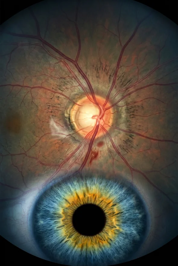
What causes a detached retina is usually the ageing of the clear gel inside the eye, which shrinks, pulls on the retina and tears it, letting fluid slip underneath. Short sight, previous cataract surgery, eye injury and family history raise the risk. New floaters, flashes of light or a dark curtain are warning signs and need same-day care. Retina specialists Doç. Dr. Muhammet Derda Özer and Op. Dr. Aysan Bayram treat detachments in Istanbul.
Sudden flashes, new floaters or a shadow over your vision? Get seen today.
The NHS, the US National Eye Institute (NEI) and the American Academy of Ophthalmology (AAO) give the same advice: go to an eye doctor or an emergency department right away. In the UK, call NHS 111. Do not wait for a routine appointment, and do not wait to travel anywhere for treatment. If you are already in Istanbul, message us and we will arrange an urgent examination with a retina surgeon: {{urgent appointment availability and how to reach the coordinator}}.
What causes a detached retina?
A retinal detachment happens when the retina, the thin light-sensing layer lining the back of the eye, is pulled or pushed away from the tissue that feeds it. A detached retina stops working, so the vision it supplies fades. Doctors recognise three types, and each has a different cause:
| Type | What causes it | Typical situation |
|---|---|---|
| Rhegmatogenous (the most common type) | A tear or hole in the retina lets fluid from the eye's gel seep underneath and lift it off, like a bubble forming under wallpaper | Ageing of the vitreous gel, short sight, previous eye surgery, injury |
| Tractional | Scar tissue on the retina's surface pulls the retina away from the back of the eye | Mostly advanced diabetic eye disease; also retinal vein occlusion and sickle cell retinopathy |
| Exudative | Fluid collects under the retina without any tear, because blood vessels or the layer beneath leak | Inflammation, infection, tumours and some vascular conditions; usually treated by treating the cause |
What is the most common cause of retinal detachment?
The most common cause is posterior vitreous detachment (PVD) leading to a retinal tear. The vitreous is the clear gel that fills the eye. With age it liquefies and shrinks, and eventually peels away from the retina. This is normal and usually harmless: according to EyeWiki, the AAO's clinical encyclopedia, PVD affects most eyes by the eighth decade of life, typically starting in the sixth or seventh decade, and earlier in short-sighted eyes or after cataract surgery. The NHS notes that it is not clear exactly why PVD leads to detachment in some people and not others.
When the gel peels away smoothly, nothing more happens. When it sticks to the retina and tugs hard enough, it makes a tear. A 2025 review in Wiener Medizinische Wochenschrift explains that a rhegmatogenous detachment needs three ingredients together: traction on the retina, a full-thickness break in it and liquefied vitreous that can flow through the break.
How often does a tear form? EyeWiki reports that people who have symptoms during a PVD have about a 15% risk of a retinal tear, and the 2025 review found that 14.3% of people attending with flashes or floaters had a tear or a detachment. When the PVD also causes bleeding into the gel, the chance of a tear is much higher: EyeWiki quotes 50–70%, against 15% without bleeding. How often does a tear become a detachment? EyeWiki states that 33–55% of symptomatic horseshoe-shaped tears with continuing traction progress to detachment, and that sealing the tear promptly with laser or freezing lowers that to under 5%. That is why a tear found early is a very different problem from a detachment found late.
Who is most at risk of a detached retina?
The NHS, NEI and AAO list the same main risk factors. The figures below come from a 2025 review (Xiong et al.) unless stated.
| Risk factor | What the evidence says |
|---|---|
| Short sight (myopia) | About a tenfold higher incidence in people with more than 3 dioptres of myopia; up to 50% of detachments in eyes with their natural lens occur in short-sighted people. Severe (degenerative) myopia is a listed NEI risk factor. |
| Cataract surgery | A risk of about 0.5–0.6% after cataract surgery, roughly ten times the general population, or about 1 in 500 operations in people over 40. Up to 44% of people presenting with a detachment have had cataract surgery. A tear in the lens capsule during surgery raises the risk further. |
| Eye injury | A serious blow or penetrating injury can tear the retina. NEI recommends safety goggles for risky activities and sport. |
| Family history or a detachment in the other eye | The NHS and AAO list both. The review reports up to a 10% risk of a detachment in the fellow eye. Inherited conditions such as Stickler and Marfan syndromes also raise the risk. |
| Weak areas of retina | Lattice degeneration (thin, weak patches) and retinoschisis are NEI risk factors; EyeWiki calls lattice degeneration the most important risk factor for rhegmatogenous detachment. |
| Age and sex | The peak is at 60–70 years, with a smaller peak at 20–30 years in highly short-sighted people. Men have about twice the risk of women. |
| Diabetic retinopathy | Proliferative diabetic retinopathy is the most common cause of tractional detachment. |
How common is it? A 2024 systematic review of 33 studies from 21 countries, summarised in the 2025 review, put the incidence of rhegmatogenous detachment at 12.17 per 100,000 people a year worldwide and 14.52 per 100,000 in Europe, and found it rising over time. EyeWiki gives a rough figure of 1 in 10,000 people. It is uncommon, but it can be treated if it is caught in time.
What are the warning signs of a detached retina?
The warning signs and symptoms of a detached retina are a sudden shower of new floaters, flashes of light, and a dark shadow or grey curtain across part of your vision. The NHS, NEI and AAO describe the same signs:
- New floaters: a lot of new dots, lines, specks or cobwebs drifting across your vision
- Flashes of light in one or both eyes, often at the edge of vision and most noticeable in the dark. EyeWiki says flashes occur in about half of people with symptomatic PVD
- A dark shadow or grey curtain spreading in from the side or across the middle of your vision
- Sudden blurring of sight
Does a detached retina hurt? No. The AAO states that a detached retina does not normally cause pain. This is why people sometimes put it off: the eye looks normal and feels normal.
Detached retina symptoms: what do they look like?
You cannot see a detached retina by looking at the eye, in a mirror or on a photograph of the outside of the eye. Only an examination with dilating drops can show it. What changes is what you see:
| What you notice | What is happening |
|---|---|
| A few new floaters and a brief flash | Often a harmless PVD, but only an examination can tell whether the retina has torn |
| A sudden "shower" of dark spots, like soot or smoke | The retina may have torn and bled into the gel |
| A curved shadow creeping in from one side, like a curtain being drawn | Part of the retina has lifted; the shadow is the area it no longer serves |
| Blurred or wavy central vision | The centre of the retina (the macula) may be involved: this is the most urgent situation |
Most people with new floaters do not have a detachment, but nobody can tell which person is which without an examination. Treat any sudden change as urgent.
How long before retinal detachment causes blindness?
There is no safe waiting time. Untreated, a retinal detachment usually leads to blindness in that eye, and the sooner it is repaired the better the result. The NEI says that waiting allows more of the retina to detach, which increases the risk of permanent vision loss or blindness. The AAO says you could quickly and permanently lose sight in the affected eye. How fast it spreads varies from person to person, so nobody can promise you days.
What matters most is the macula, the small central area of the retina you use for reading and recognising faces:
| Situation | What it means | Outlook (2025 review) |
|---|---|---|
| Macula-on: the centre is still attached | Central vision may still be good; this is an emergency to stop the detachment reaching the centre | About 90% reach 20/40 vision or better |
| Macula-off: the centre has detached | Central vision is already blurred or lost | About 50% reach 20/50 or better; results are worse if the macula has been detached for a week or more |
Timing matters even after the macula comes off. A 2023 study of 719 eyes in Japan (Miyake et al., Ophthalmology Retina), summarised by the AAO, found that vision at 6 months was significantly better when surgery was done within 2 days of the macula detaching than at 3 days, and better at 3 days than at 4 or more. The authors noted that this has to be balanced against the availability of an experienced surgical team.
What should you do if you have symptoms?
Go to an eye emergency service or an emergency department today
In the UK, call NHS 111 or go to an eye casualty. Say you have new floaters or flashes or a shadow, so you are seen quickly.
Do not wait to see if it settles,
and do not wait until a flight, a holiday or a weekend is over.
Expect a dilated examination
The doctor puts drops in to widen the pupil and looks at the whole retina with a special lens. NEI notes that ultrasound or OCT can add detail, for example if bleeding blocks the view.
If a tear is found without a detachment,
laser or freezing treatment can usually seal it in the clinic. The NEI says small tears can be treated in the office, while larger detached areas need surgery in a hospital.
If your exam is normal, you still need to know the signs
EyeWiki advises a follow-up dilated examination about a month after a symptomatic PVD, because tears can develop later. Come back at once if your symptoms change.
How is a detached retina treated? Urgent surgery explained
A detached retina is repaired with surgery that seals the tear and holds the retina in place until it heals. The NHS says surgery will usually stop your vision getting worse. There are three main operations, and your surgeon chooses according to where the tear is, how much retina is detached, your age and whether you have had cataract surgery.
| Operation | How it works | What to know |
|---|---|---|
| Vitrectomy | Keyhole surgery removes the vitreous gel, the tear is sealed with laser or freezing, and the eye is filled with a gas bubble, air or silicone oil that acts as a splint | The gas bubble dissolves by itself; silicone oil is removed in a later operation (AAO). You may need to keep your head in a set position for up to a week (Moorfields). |
| Scleral buckle | A silicone band is stitched to the outside of the eye wall to push the wall towards the tear | Usually left in place permanently |
| Pneumatic retinopexy | A gas bubble is injected and you hold a specific head position so it presses the retina back; the tear is sealed with laser or freezing | Suits selected detachments. In the PIVOT trial, 80.8% were reattached after one operation at 12 months, against 93.2% for vitrectomy |
How successful is retinal detachment surgery? The NEI says treatment is successful for about 9 out of 10 people, and EyeWiki reports anatomical success above 90% with modern techniques. The NHS reminds patients that a detachment can come back, and the AAO says about 1 in 20 people need a second operation. Possible complications include infection, bleeding, a rise in eye pressure leading to glaucoma, and cataract.
What is recovery like?
- 2 to 6 weeks of blurred vision, a sore or red eye, time off work and no driving, according to the NHS
- Vision typically starts to improve 4–6 weeks after surgery, and healing can continue for a year or more (AAO)
- No flying, no high altitude and no scuba diving while a gas bubble is in the eye. Changes in altitude make the gas expand and can dangerously raise eye pressure (AAO). Moorfields gives these maximum times: air up to 2 weeks, SF6 gas up to 4 weeks, C2F6 gas up to 8 weeks, C3F8 gas up to 12 weeks; with silicone oil, no flying for two weeks unless your surgeon clears you
If you are an international patient, this is why the order matters: get the detachment repaired first, wherever you are, and plan any journey with your surgeon afterwards.
Can a detached retina be prevented?
Often it cannot. The NEI says that because detachment is often caused by ageing, there is often no way to prevent it. What you can do:
- Know the warning signs and act on them the same day. This is the most useful step, especially if you are short-sighted, have had cataract surgery or have had a detachment in the other eye.
- Wear safety goggles or protective eyewear for racquet sports, DIY and other risky activities, because injury is a preventable cause.
- Control diabetes and attend your retinal screening, because tractional detachment comes from advanced diabetic eye disease.
- Tell your doctor about family history or inherited conditions that affect the eye.
- Have tears treated early. Sealing a symptomatic tear lowers the chance of detachment to under 5% (EyeWiki).
There are no proven natural remedies, drops or supplements that prevent or reverse a detachment. Anything that delays an examination is a risk.

Retinal detachment care at Laser Istanbul Clinic
Retinal detachments and other retina conditions are treated by Doç. Dr. Muhammet Derda Özer (Doç. = Associate Professor), who treats retinal disease alongside cornea and uveitis and speaks Turkish and English, and Op. Dr. Aysan Bayram, a specialist in cataract surgery, laser and retinal disease who speaks English. Operations take place in a fully equipped, Ministry of Health–licensed operating theatre in Istanbul. Your examination and scans, surgery by your named surgeon, medication, follow-up checks in Istanbul, a personal interpreter and remote follow-up are all part of your care plan.
Because a fresh detachment is an emergency, we do not ask anyone with symptoms to wait for a flight. If you are in Istanbul or can reach it safely, we arrange an urgent examination. Otherwise, go to your nearest eye emergency service first. We can also review your scans and give a second opinion on a repair that has already been done, or on a planned operation.
Frequently asked questions
Most detachments start when the vitreous gel in the eye shrinks with age, tugs on the retina and makes a tear. Fluid then passes through the tear and lifts the retina. Less common causes are scar tissue pulling on the retina (often from diabetic eye disease) and fluid leaking under the retina from inflammation, infection or tumours.
A tear in the retina caused by posterior vitreous detachment, the normal age-related separation of the gel from the retina. It usually starts in the sixth or seventh decade of life, earlier in short-sighted eyes or after cataract surgery. Most PVDs never cause a tear, but about 1 in 7 people who attend with flashes or floaters has a tear or detachment.
A sudden shower of new floaters, flashes of light, a dark shadow or grey curtain across part of your vision, or sudden blurring. The condition is painless. If you notice any of these, see an eye doctor or go to an emergency department the same day.
There is no fixed timetable, and it varies from person to person, so treat it as an emergency. Untreated, it usually leads to blindness in that eye. If the macula is still attached, the best chance of keeping good central vision is repair before the detachment reaches the centre. If the macula has detached, a large study found better vision when surgery was done within 2 days than later.
No, retinal detachment needs treatment. The NHS and NEI describe surgery as the standard treatment, and without it the detachment usually leads to blindness in that eye. A small tear without detachment can often be sealed with laser or freezing.
The detachment itself is painless, and the NHS says surgery is usually done under local anaesthetic as an outpatient procedure. Afterwards the eye is often sore and red for some days to weeks. Your surgeon will tell you which drops and painkillers to use.
Yes. A review of the evidence reports up to a 10% risk of detachment in the other eye, and a detachment in one eye is a listed risk factor for the other. That is why people who have had one detachment should know the warning signs for life and see a doctor straight away if they appear.
No. With an air or gas bubble you must not fly, travel to high altitudes or scuba dive until your surgeon confirms that the bubble has gone. Depending on the gas, that can take 2 to 12 weeks, and your surgeon will tell you your own date.
Cataract surgery is a recognised risk factor: about 0.5–0.6% of people develop a detachment after it, roughly 10 times the general population, and the risk is higher if the lens capsule tears during surgery. Short sight raises the risk further. Ask your surgeon about your own risk factors before any eye operation.
Worried about your eyes, or planning treatment?
Send your scans and symptoms and one of our retina surgeons reviews them, tells you honestly what they see and what the next step is. For emergency symptoms, go to the nearest eye emergency service first.
Sources for the facts on this page
- All sources opened and checked on 7 Oct 2026.
- NHS. Detached retina (retinal detachment), last reviewed 4 Aug 2023. https://www.nhs.uk/conditions/detached-retina-retinal-detachment/
- National Eye Institute (NIH). Retinal Detachment. https://www.nei.nih.gov/learn-about-eye-health/eye-conditions-and-diseases/retinal-detachment
- American Academy of Ophthalmology. Detached Retina. https://www.aao.org/eye-health/diseases/detached-torn-retina
- EyeWiki (AAO). Retinal Detachment. https://eyewiki.org/Retinal_Detachment
- EyeWiki (AAO). Posterior Vitreous Detachment. https://eyewiki.org/Posterior_Vitreous_Detachment
- EyeWiki (AAO). Horseshoe or Flap Tear. https://eyewiki.org/Horseshoe_or_Flap_Tear
- EyeWiki (AAO). Exudative Retinal Detachment. https://eyewiki.org/Exudative_Retinal_Detachment
- Xiong J, Tran T, Waldstein SM, Fung AT. A review of rhegmatogenous retinal detachment: past, present and future. Wiener Medizinische Wochenschrift 2025;175(7-8):186-202. https://pmc.ncbi.nlm.nih.gov/articles/PMC12031774/
- Moorfields Eye Hospital NHS Foundation Trust. Retinal detachment: diagnosis and treatment. https://www.moorfields.nhs.uk/eye-conditions/retinal-detachment/diagnosis-and-treatment
- American Academy of Ophthalmology. Macula-off retinal detachments fare best when corrected within 2-3 days (summary of Miyake et al., Ophthalmology Retina 2023). https://www.aao.org/education/editors-choice/macula-off-retinal-detachments-fair-best-when-corr