
- Home
- Knowledge centre
- Retina & glaucoma guides
- Diabetic Retinopathy: Treatment and Prevention
Diabetic Retinopathy: Treatment and Prevention
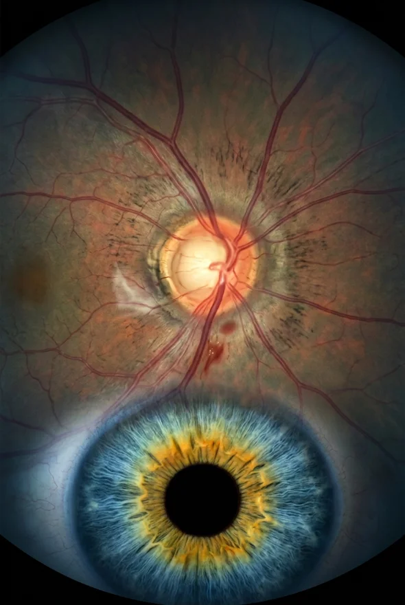
Diabetic retinopathy is damage to the retina's blood vessels caused by long-term high blood sugar. It often causes no symptoms at first, so regular screening is the main safeguard. Early stages are watched; advanced disease is treated with anti-VEGF injections, laser, steroid implants or vitrectomy surgery. Keeping blood sugar, blood pressure and cholesterol under control lowers the risk. In Istanbul, retina surgery is led by Doç. Dr. (Associate Professor) Muhammet Derda Özer and Op. Dr. Aysan Bayram.
What is diabetic retinopathy, and what causes it?
Diabetic retinopathy is an eye condition that can cause vision loss and blindness in people who have diabetes. It affects the blood vessels in the retina, the light-sensitive layer at the back of the eye, according to the US National Eye Institute (NEI). Anyone with any type of diabetes can develop it, including type 1, type 2 and gestational diabetes.
Diabetic retinopathy causes. The cause is long-term high blood sugar. The NHS explains that high glucose damages the retinal blood vessels, and that the risk rises with the length of time you have had diabetes, high blood pressure and high cholesterol. Other risk factors named by EyeWiki, the clinical encyclopedia of the American Academy of Ophthalmology (AAO), are pregnancy, smoking and ethnicity.
How it happens, step by step (pathophysiology). In plain language, the sequence is:
- Chronic high blood sugar causes inflammation and oxidative stress in the retinal capillaries.
- The cells that support the vessel walls (pericytes) and the capillaries themselves are damaged.
- Damaged vessels leak fluid or close off, so parts of the retina receive less blood (ischaemia).
- The starved retina releases more of a growth signal called VEGF (vascular endothelial growth factor).
- VEGF makes vessels leakier, which swells the macula (diabetic macular oedema), and triggers new, abnormal vessels to grow (neovascularisation).
- The new vessels are fragile: they bleed into the eye's gel (vitreous haemorrhage) and form scar tissue that can pull the retina off the back of the eye (tractional retinal detachment).
How common is it? A 2021 meta-analysis of 59 population-based studies (Teo et al., Ophthalmology) found diabetic retinopathy in about 22% of people with diabetes worldwide (22.27%), and vision-threatening disease in about 6%. It estimated 103 million people with the condition in 2020, rising to about 160 million by 2045. The NEI says that over time more than half of people with diabetes will develop some diabetic retinopathy, and about 1 in 15 will develop diabetic macular oedema. The World Health Organization (WHO) reports that the number of people living with diabetes rose from 200 million in 1990 to 830 million in 2022, so the burden is growing.
What are the symptoms of diabetic retinopathy?
Early diabetic retinopathy usually has no symptoms. The NHS, NEI and AAO all say that the early stages can be present without any change you can notice. That is why you can have vision-threatening disease and still read the newspaper comfortably.
Early stage diabetes eyes symptoms: what you can and cannot feel
In the early stage, most people feel nothing at all. Some notice vision that is a little blurry or that changes from day to day, but this is not specific to retinopathy and is not a reliable warning sign. Do not wait for symptoms: attend every screening appointment, even if your eyesight seems fine.
Symptoms of diabetic retinopathy in later stages
As the disease advances, the NHS, NEI and AAO list:
- blurred or fluctuating vision
- trouble reading or seeing things in the distance
- floaters: dark spots, dots, lines or cobweb-like streaks in your vision
- dark or blank areas in your vision
- reduced night vision and colours that look faded
- loss of vision
Both eyes can be affected, although not always to the same degree.
When is it urgent? Dark floating spots or streaks can mean the retina is bleeding. The NEI says it is important to get treatment right away if you notice them, because without treatment scars can form. Scar tissue can also pull on the retina and detach it. If you see a sudden shower of floaters, flashes of light or a shadow across your vision, read Retinal detachment: warning signs and urgent surgery and contact an eye emergency service the same day.
What are the 5 stages of diabetic retinopathy?
Diabetic retinopathy is graded in five stages on the international clinical scale. This scale, proposed in 2003 by an international group of 31 experts from 16 countries, has three low-risk stages, then severe non-proliferative retinopathy, then proliferative retinopathy. Doctors call this the classification of diabetic retinopathy; the table turns it into plain language.
| Stage | What the doctor sees | Usual symptoms | Usual approach |
|---|---|---|---|
| 1. No apparent retinopathy | A normal retina | None | Keep to your screening schedule |
| 2. Mild non-proliferative (NPDR) | A few microaneurysms (tiny bulges in the vessel walls) | None | Monitoring and diabetes control; the NHS says no treatment is needed in the early stages |
| 3. Moderate NPDR | More microaneurysms, small dot-and-blot haemorrhages, sometimes cotton-wool spots | Often none | Closer monitoring; swelling at the macula is treated if it develops |
| 4. Severe NPDR | The "4-2-1 rule": bleeding and microaneurysms in all 4 quadrants, or venous beading in 2 or more, or abnormal vessel branching (IRMA) in 1 or more | Often none | Frequent specialist review; treatment may be considered |
| 5. Proliferative (PDR) | New abnormal vessels, bleeding into the eye, scar tissue | Floaters, sudden blur or loss of vision | Laser, anti-VEGF injections, and sometimes vitrectomy |
Diabetic macular oedema (DME) is graded separately because it can occur at any stage. It means fluid has collected in the macula, the central part of the retina. The AAO calls macular oedema the leading cause of vision loss in people with diabetes. In the UK the NHS describes three stages (background, pre-proliferative and proliferative retinopathy), which roughly correspond to the stages above.
How is diabetic retinopathy diagnosed, and how often should you be screened?
The standard test is a dilated eye examination. Drops widen your pupils so the doctor, or a camera, can see the whole retina. The NHS describes screening as reading a letter chart, then dilating drops, then photographs of the back of the eye, in an appointment of about 30 minutes; your vision may be blurred for a few hours afterwards, so you should not drive. If severe disease or macular oedema is suspected, the NEI says doctors use fluorescein angiography (a dye test) to see leaking vessels, and EyeWiki adds that an optical coherence tomography (OCT) scan shows swelling in cross-section.
Recommended screening intervals:
| Who | How often | Source |
|---|---|---|
| Adults with type 2 diabetes | At diagnosis, then every year | AAO Preferred Practice Pattern, 2024 |
| People with type 1 diabetes | Every year, starting 5 years after diagnosis | AAO, 2024 |
| People with diabetes aged 12 or over in the UK | Every year, or every 2 years if the last two tests found no changes | NHS |
| Pregnant women with diabetes | An eye exam as early as possible, then as advised; progression can be rapid | AAO, NEI |
| Anyone who already has diabetic retinopathy | As often as every 2 to 4 months | NEI |
The AAO notes that only about 60% of people with diabetes have the recommended yearly screening. Missing an appointment is the most avoidable mistake in diabetic eye care.
Diabetic retinopathy treatment: what works at each stage
Treatment depends on the stage and on whether the macula is swollen. The NHS says that in the early stages (background and pre-proliferative) you will not need treatment and will be monitored at your regular screening appointments. For advanced disease, the NHS, NEI and AAO describe four options: injections, laser, steroid implants and surgery. Your specialist advises which suits you.
| Treatment | What it does | Typically used for |
|---|---|---|
| Anti-VEGF injections | Block VEGF, so vessels leak less and abnormal vessels regress | Diabetic macular oedema; proliferative disease; sometimes before surgery |
| Laser (panretinal photocoagulation, PRP) | Many tiny burns across the outer retina shrink abnormal vessels | Proliferative disease |
| Focal laser | Seals leaking vessels near the macula | Selected macular oedema away from the centre |
| Steroid implants or injections | Release steroid slowly to reduce swelling | Macular oedema |
| Vitrectomy | Removes blood and scar tissue from inside the eye | Heavy bleeding, extensive scarring, tractional detachment |
Anti-VEGF injections
Anti-VEGF drugs are the first-line treatment for diabetic macular oedema, and they can also treat proliferative disease. The NEI says they can slow down or even reverse diabetic retinopathy. The medicine is injected into the eye after numbing drops, and the AAO stresses that povidone iodine should be used for every injection, because skipping it has been reported to carry an unacceptably high risk of infection (endophthalmitis). According to EyeWiki, the typical schedule for macular oedema is six monthly injections at first, then perhaps two to four in the second year and minimal treatment after that.
Evidence from the US Diabetic Retinopathy Clinical Research Network (DRCR) supports this. In Protocol T, aflibercept gave better results than bevacizumab or ranibizumab when vision was 20/50 or worse, and Protocol V showed that watching eyes with good vision (20/25 or better) was comparable to treating immediately. For earlier disease, Protocol W gave aflibercept or a sham injection to 328 people with moderate to severe NPDR and no central swelling. After 4 years, 33.9% of the aflibercept group had developed proliferative disease or sight-threatening macular oedema, compared with 56.9% of the sham group. Average vision was the same in both groups, so the trade-off is repeated injections for a lower risk of complications, not better sight. That decision is for you and your specialist.
Laser treatment (panretinal photocoagulation)
PRP remains an important treatment for proliferative diabetic retinopathy. The Diabetic Retinopathy Study, which enrolled over 1,700 patients, found that PRP cut the risk of severe vision loss by more than half. At 4 years, 44% of untreated high-risk eyes had severe vision loss, against 20% of treated eyes (EyeWiki). Treatment is typically 1,500 to 5,000 laser burns over 1 to 4 sessions. The cost is that PRP can permanently reduce peripheral, colour and night vision.
The most useful trial for choosing between laser and injections is DRCR Protocol S. In 394 eyes with proliferative disease, vision at 5 years averaged 20/25 in both the ranibizumab group and the laser group. The injection group received about 19 injections over the five years; the laser group had about 5 laser treatments. Vision-impairing macular oedema developed in 22% of the injection group and 38% of the laser group, and visual field loss was less severe with injections. The authors concluded that either approach is reasonable and that individual patient factors should guide the choice. One practical factor: injections work only if you can keep coming back, while laser is completed in a few visits.
Steroid implants and injections
Steroid medicines, given as an implant or injection, reduce swelling in the macula. The NHS lists steroid-releasing implants among the standard options for diabetic eye disease, and the NEI mentions corticosteroid injections. According to EyeWiki, a 0.7 mg dexamethasone implant lasts about 3 to 6 months and a 0.19 mg fluocinolone implant about 3 years.
Vitrectomy for advanced diabetic eye disease
A vitrectomy is keyhole surgery that removes blood and scar tissue from inside the eye. The NEI says it may be recommended if the retina is bleeding a lot or there is a lot of scarring. EyeWiki lists vitrectomy for advanced disease with haemorrhage or tractional retinal detachment, and notes that anti-VEGF is sometimes given beforehand to reduce the risk of bleeding. In Istanbul, planned vitrectomy is performed by Doç. Dr. Özer or Op. Dr. Bayram in a fully equipped, Ministry of Health–licensed operating theatre. If a gas bubble is placed in the eye, you cannot fly until it has gone, so plan your stay with your surgeon. For recovery questions, see Blurry vision after retina surgery: what is normal.
Can diabetic retinopathy be reversed?
Treatment can stop vision getting worse, but it cannot undo damage that has already happened. The NEI says treatment won't undo damage to your vision but can stop it worsening. Some changes can improve: the NEI notes that anti-VEGF drugs can slow or reverse retinopathy, and the AAO says good sugar control can bring some vision back. The earlier the problem is found, the more vision there is to protect.

Diabetic retinopathy treatment with eye drops: do they work?
No eye drop is a recognised treatment for diabetic retinopathy. The treatments listed by the NHS, NEI and AAO are injections, laser, implants and surgery, together with control of blood sugar, blood pressure and cholesterol. None of them is an eye drop. Be cautious about products sold online as "natural" cures or drops: if one claims to reverse retinopathy, ask your eye doctor before using it, and never replace screening or prescribed treatment with it. Eye drops do have a role elsewhere: dilating drops are used for the examination, and your surgeon may prescribe drops after an injection or an operation.
How to prevent eye damage from diabetes
The best prevention is to keep blood sugar, blood pressure and cholesterol in range and to attend every screening. The NEI puts it simply: you can lower your risk of diabetic retinopathy by controlling your diabetes. The evidence is strong:
- Blood sugar. In the Diabetes Control and Complications Trial (1,441 people with type 1 diabetes, 6.5 years), intensive treatment reduced the risk of developing retinopathy by 76% and slowed progression by 54% in people who already had mild retinopathy. In the UK Prospective Diabetes Study (type 2), each 1% reduction in HbA1c was associated with a 37% reduction in microvascular complications (EyeWiki). A common target is an HbA1c below 7%, but your own target should be agreed with your diabetes team.
- Blood pressure and cholesterol. The NHS and NEI name high blood pressure and high cholesterol as risk factors, and the AAO names high blood pressure and kidney problems, so treating them protects the eyes as well as the heart and kidneys.
- Fenofibrate. In the ACCORD Eye trial, adding fenofibrate to simvastatin lowered the odds of retinopathy progression by about 40% over 4 years (EyeWiki). This is a decision for your doctor, not something to start on your own.
- No smoking. The NHS lists avoiding smoking among its prevention advice.
- Activity and diet. The NEI recommends regular physical activity, healthy eating and taking your medication as prescribed. WHO advises 150 minutes of moderate exercise a week for preventing type 2 diabetes.
- Screening and fast action. Attend every appointment and report any change in vision at once.
- Pregnancy. If you have diabetes and are pregnant, the NEI advises a comprehensive dilated eye exam as soon as possible.
A caution on rapid sugar lowering. The 2024 AAO guideline notes that rapid blood-sugar control with newer agents such as semaglutide can accelerate the onset of retinopathy in some people. This is no reason to stop treatment. It is a reason to have your eyes checked around any big change in your diabetes treatment, and to tell your diabetes team if you already have retinopathy.

When does it make sense to travel to Istanbul for diabetic eye care?
Planned surgery and laser suit travel; repeated injections and yearly screening are best done close to home. If you have a non-clearing haemorrhage, diabetic scar tissue or want a second opinion, a vitrectomy can be scheduled in advance, your scans can be reviewed online first, and you can plan your stay around the surgeon's advice. Anti-VEGF injections are given again and again, so having them near your home makes more sense, and your yearly screening should stay with your local service.
Retina and diabetic eye disease are led by Doç. Dr. Muhammet Derda Özer, who also treats cornea, uveitis and ocular tumours and speaks Turkish and English, and Op. Dr. Aysan Bayram, a female surgeon who speaks English and operates on cataract, laser and retinal disease. Before any operation your surgeon asks about your diabetes control and medicines, because they affect planning.
Do not travel if you have sudden new symptoms such as a curtain across your vision or a sudden shower of floaters: get seen by an eye emergency service near you at once.
What the Istanbul price includes. Vitrectomy starts from {{£ price}} per eye. It includes the pre-operative examination and scans, surgery by the named surgeon, medication, follow-up checks in Istanbul, a personal interpreter, a coordinator and remote follow-up. Hotel and airport transfers are not included; they are arranged on request for a separate charge, and flights are not included.
Frequently asked questions
Diabetic retinopathy is damage to the retina's blood vessels caused by long-term high blood sugar. It starts silently, may cause leaking, bleeding and abnormal vessel growth, and can lead to vision loss if untreated. Regular screening finds it before you notice symptoms.
There are usually none. If symptoms appear, they include blurred vision, floaters, dark patches and trouble seeing at night. Because early disease is silent, yearly screening matters more than waiting for signs.
No apparent retinopathy, mild non-proliferative, moderate non-proliferative, severe non-proliferative and proliferative retinopathy. Diabetic macular oedema is graded separately. The first stages are monitored; the later ones are treated.
Early stages are watched and managed with better diabetes control. Advanced disease is treated with anti-VEGF injections, laser, steroid implants or vitrectomy, chosen according to the stage and whether the macula is swollen. Your specialist explains the options and risks.
No eye drop is a standard treatment. The recognised treatments are injections, laser, implants, surgery and control of blood sugar, blood pressure and cholesterol. Drops are used for examinations and may be prescribed after procedures.
It can be controlled, but damage already done cannot be undone. Treatment aims to stop vision getting worse, and good control of diabetes and anti-VEGF treatment can improve some changes. Finding it early gives the best result.
At least once a year. In the UK you are invited every year, or every 2 years if your last two tests showed no changes. If you already have retinopathy, you may need checks as often as every 2 to 4 months.
Screening in the NHS programme is by invitation. For treatment in Istanbul, vitrectomy starts from {{£ price}} per eye, with the pre-operative examination and scans, surgery, follow-up and interpreter included; hotel and transfers are extra. Your surgeon confirms the final price in writing after reviewing your scans.
Often yes for planned surgery, after your surgeon reviews your scans and your diabetes control. Send your reports through the free online consultation and your surgeon will tell you honestly whether travel makes sense.
Worried about diabetes and your eyes?
Upload your latest eye reports and scans and a retina surgeon at Laser Istanbul Clinic tells you whether treatment is needed, what it involves and whether it makes sense to travel. Retina care is led by Doç. Dr. Muhammet Derda Özer and Op. Dr. Aysan Bayram.
Sources for the facts on this page
- NHS. Diabetic retinopathy. https://www.nhs.uk/conditions/diabetic-retinopathy/
- NHS. Diabetic retinopathy: treatment. https://www.nhs.uk/conditions/diabetic-retinopathy/treatment/
- NHS. Diabetic eye screening. https://www.nhs.uk/conditions/diabetic-eye-screening/
- National Eye Institute (NIH). Diabetic Retinopathy. https://www.nei.nih.gov/learn-about-eye-health/eye-conditions-and-diseases/diabetic-retinopathy
- American Academy of Ophthalmology. What Is Diabetic Retinopathy? https://www.aao.org/eye-health/diseases/what-is-diabetic-retinopathy
- American Academy of Ophthalmology. Diabetic Retinopathy Preferred Practice Pattern (2024). https://www.aao.org/education/preferred-practice-pattern/diabetic-retinopathy-ppp
- EyeWiki (AAO). Diabetic Retinopathy. https://eyewiki.org/Diabetic_Retinopathy
- EyeWiki (AAO). Diabetic Macular Edema. https://eyewiki.org/Diabetic_Macular_Edema
- EyeWiki (AAO). Panretinal Photocoagulation. https://eyewiki.org/Panretinal_Photocoagulation
- Teo ZL, et al. Global Prevalence of Diabetic Retinopathy and Projection of Burden through 2045. Ophthalmology 2021. https://pubmed.ncbi.nlm.nih.gov/33940045/
- Wilkinson CP, et al. Proposed international clinical diabetic retinopathy and diabetic macular edema disease severity scales. Ophthalmology 2003. https://pubmed.ncbi.nlm.nih.gov/13129861/
- Gross JG, et al. Five-Year Outcomes of Panretinal Photocoagulation vs Intravitreous Ranibizumab for Proliferative Diabetic Retinopathy (DRCR Protocol S). JAMA Ophthalmology 2018. https://pmc.ncbi.nlm.nih.gov/articles/PMC6233839/
- Maturi RK, et al. Four-Year Visual Outcomes in the Protocol W Randomized Trial of Intravitreous Aflibercept. JAMA 2023. https://pmc.ncbi.nlm.nih.gov/articles/PMC10408259/
- Diabetes Control and Complications Trial Research Group. The effect of intensive treatment of diabetes on the development and progression of long-term complications in insulin-dependent diabetes mellitus. N Engl J Med 1993. https://pubmed.ncbi.nlm.nih.gov/8366922/
- ACCORD Study Group and ACCORD Eye Study Group. Effects of medical therapies on retinopathy progression in type 2 diabetes. N Engl J Med 2010. https://pmc.ncbi.nlm.nih.gov/articles/PMC4026164/ (the 40% figure as summarised in EyeWiki, source 7)
- World Health Organization. Diabetes fact sheet. https://www.who.int/news-room/fact-sheets/detail/diabetes