
- Home
- Knowledge centre
- Eye conditions & symptoms A–Z
- What Is the Macula? Macular Degeneration (Yellow Spot): Symptoms and Treatment
What Is the Macula? Macular Degeneration (Yellow Spot): Symptoms and Treatment
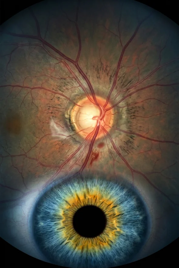
The macula is the small central part of the retina that gives you sharp, straight-ahead vision for reading, faces and driving. In age-related macular degeneration (AMD, the "yellow spot disease") it wears out, blurring or distorting the centre of your sight while side vision stays. About 80% of cases are the slow dry form and 20% the faster wet form. Wet AMD can be treated with eye injections; dry AMD has no cure, but supplements, lifestyle and new therapies can slow it. Our retina surgeon, Doç. Dr. Muhammet Derda Özer, assesses retinal disease.
What is the macula, and what does "macula" mean?
The macula is the part of the retina that controls sharp, straight-ahead vision, according to the US National Eye Institute (NEI). The retina is the light-sensitive layer lining the back of the eye, and the macula sits at its centre. It is the area you use to read a page, recognise a face, thread a needle or read a road sign.
The word "macula" is Latin for "spot". The full anatomical name, macula lutea, means "yellow spot", which is why people searching in other languages, and many older patient leaflets, call macular degeneration the "yellow spot disease". The macula looks yellowish under an eye doctor's instrument because it contains yellow pigments (lutein and zeaxanthin), the same two substances that appear in the supplements discussed below.
Here is a simple way to picture it. Your retina is like the sensor in a camera, and the macula is its sharpest, most detailed patch. The rest of the retina gives you side (peripheral) vision, which helps you move around but cannot read small print. When the macula is damaged, the centre of what you look at is affected first.
What is macular degeneration (yellow spot disease)?
Macular degeneration is a condition in which the macula gradually deteriorates. The usual form is age-related macular degeneration (AMD), which the NHS describes as a common condition that affects the middle part of your vision and typically starts in a person's 50s or later. It does not cause complete blindness: the NEI states that AMD doesn't cause complete blindness, but losing central vision can make it harder to see faces, read, drive or do close-up work. AMD is painless and does not change how your eyes look.
How common is it? A 2014 meta-analysis in The Lancet Global Health (129,664 people, 39 studies) estimated that 8.69% of people aged 45 to 85 have some form of AMD, mostly early disease, and 0.37% have late AMD. It projected 196 million people with AMD worldwide in 2020, rising to 288 million by 2040. Prevalence was higher in people of European ancestry than in Asian or African populations.
There are two forms:
| Dry AMD (atrophic) | Wet AMD (neovascular) | |
|---|---|---|
| Share of cases | About 80% (AAO) | About 20% (AAO) |
| What happens | The macula thins with age and yellow deposits called drusen build up | Abnormal new blood vessels grow under the retina and leak, which can scar the macula |
| Speed | Usually gradual, over years | Much faster than dry AMD; the NHS says vision can drop within weeks or months |
| Treatment | No cure; slow it with supplements, lifestyle and, in late stages, new injections | Anti-VEGF injections; sometimes light or laser treatment |
The NEI divides dry AMD into early (no symptoms), intermediate (mild symptoms possible) and late (vision loss obvious). Anyone with AMD should know the warning signs below, because dry AMD can be followed by the wet form.
What are the early warning signs of macular degeneration?
In early dry AMD there are usually no symptoms at all, so it is often found at a routine eye test, as the NHS notes. When symptoms do appear, the main early warning signs of macular degeneration are:
- a blurred or distorted area in the centre of your vision (NHS)
- straight lines looking wavy or crooked, for example door frames, tiles or lines of text (NHS, NEI)
- blurry, dim or empty patches in the middle of your view, while the edges stay clear (NEI)
- trouble reading or recognising faces: the American Academy of Ophthalmology (AAO) compares it to looking at a clock and seeing the numbers but not the hands
- sensitivity to bright light and flickering or flashing lights (NHS)
- seeing shapes or images that are not there (hallucinations), which the NHS lists as a possible symptom of AMD
What are the first signs of wet macular degeneration?
The first signs of wet macular degeneration are the same distortion symptoms, but they appear or worsen faster: lines that suddenly look bent, a new dark or blurry spot in the middle of your sight, or a noticeable drop in central vision over days or weeks. Because the leaking vessels can scar the macula, speed matters. Anyone who notices a sudden change should be seen urgently (see "When is macular degeneration an emergency?" below).
What are the 1st signs of macular degeneration in the dry form?
The first signs of dry macular degeneration are subtle and slow: needing more light to read, print looking a little blurry or patchy, and faces taking longer to recognise. Because the change is gradual and the two eyes often change at different speeds, many people do not notice it until one eye is clearly worse.
Dry macular degeneration symptoms by stage
- Early
Usually nothing; found on a dilated eye exam
- Intermediate
Mild symptoms are possible; often still nothing
- Late
Loss of the central vision you need to see details straight ahead; blurry or wavy areas; straight lines look crooked

Macular degeneration symptoms test: the Amsler grid
The AAO recommends checking your vision with an Amsler grid, a printed square of straight lines with a dot in the middle, if you have AMD. It is a quick home check for the early warning signs, not a diagnosis.
- Hold the grid about 12 to 15 inches (30 to 38 cm) from your face, in good light.
- Cover one eye.
- Look steadily at the dot in the middle.
- Note whether any lines look wavy, bent, blurry or dim, or whether any parts of the grid are missing.
- Repeat with the other eye.
The AAO recommends testing daily and says to call your ophthalmologist right away if any lines or parts of the grid look wavy, blurry or dim. A normal result does not rule out AMD, because early disease can be silent, and regular eye tests matter even without symptoms.
What causes macular degeneration? Risk factors
The exact cause of AMD is unknown, according to the NHS, but several factors raise the risk. The NHS lists ageing, smoking, high blood pressure, being overweight and a family history of AMD. The NEI adds that risk rises from age 55 and is higher in people who are Caucasian.
| Risk factor | What it means in practice |
|---|---|
| Age | The strongest factor; AMD usually appears after 50 and risk climbs from 55 (NHS, NEI) |
| Family history | If a relative has AMD, your own risk is higher (NHS, NEI) |
| Smoking | One of the main risks you can change; both the NHS and NEI list it, and the NEI advises quitting |
| High blood pressure, overweight | Listed by the NHS; the NEI recommends keeping blood pressure and cholesterol healthy and exercising regularly |
| Ethnicity | The NEI lists being Caucasian as a higher risk; the Lancet analysis found higher prevalence in Europeans |
Having risk factors does not mean you will develop AMD, and many people with several of them keep good vision. The point is that smoking and general health are the factors you can influence.

How is macular degeneration diagnosed?
An eye doctor diagnoses AMD with a dilated eye examination, in which drops widen your pupil so the back of the eye can be inspected, and with optical coherence tomography (OCT), a painless scan that makes a cross-section picture of the macula (NEI). If you notice changes in the UK, the NHS advises seeing an optician, because early diagnosis and treatment may help stop your vision getting worse.
If you already have scans and a letter, you can send them for a second opinion: Free online consultation (upload prescription & scans) →.
Not every blur in the centre of your vision is AMD. Cataract, an out-of-date glasses prescription or astigmatism can also blur vision, which is one reason a proper examination, not self-diagnosis, is needed.
What is the best treatment for macular degeneration?
The best treatment for macular degeneration depends on the type. Wet AMD has effective medicines; dry AMD has options to slow its progression.
Wet AMD: anti-VEGF injections
For wet AMD, the main treatment is anti-VEGF injections: medicine given directly into the eye that reduces leaking from the abnormal blood vessels (NHS, AAO). The medicine is delivered through a very thin needle (AAO). Sometimes a light treatment (photodynamic therapy) or laser is used to destroy the abnormal vessels (NHS, NEI, AAO).
The evidence is strong. A Cochrane review of 16 randomised trials (6,347 people) found that anti-VEGF drugs improve vision compared with control, with ocular inflammation and raised eye pressure the most frequently reported serious eye side effects. In the large US CATT trial (1,208 patients), two common drugs, ranibizumab and bevacizumab, gave similar vision gains when given monthly: about 8 letters on the eye chart. The injections are repeated, often monthly at first. Newer drugs may allow longer gaps: in one 2026 real-world study of faricimab, 67.0% of patients reached intervals of 12 weeks or more after four monthly starter doses.
Dry AMD: slowing it down
- Supplements. For intermediate dry AMD, a vitamin and mineral formula can slow progression. The first AREDS trial (3,640 people) found that antioxidants plus zinc lowered the odds of advanced AMD (odds ratio 0.72). The AAO describes the AREDS2 formula as vitamin C 500 mg, vitamin E 400 IU, lutein 10 mg, zeaxanthin 2 mg, zinc 80 mg and copper 2 mg, and notes that smokers should avoid beta carotene because of the lung cancer risk. In the AREDS2 trial (4,203 people), adding lutein and zeaxanthin or omega-3 fatty acids did not significantly lower five-year progression to advanced AMD (31% on placebo, 29% lutein and zeaxanthin, 31% omega-3, 30% both). Supplements are for people already diagnosed with intermediate disease, and should be discussed with your eye doctor first; they are not a way to prevent AMD in healthy eyes.
- Geographic atrophy injections. Late dry AMD with patches of lost tissue is called geographic atrophy. Medicines injected into the eye may slow how fast it grows (AAO). Two of them, pegcetacoplan and avacincaptad pegol, are currently the only therapies approved by the US FDA for it. In long-term trial follow-up, monthly pegcetacoplan reduced the growth rate of subfoveal geographic atrophy by 21% over 36 months. These medicines slow the loss; they do not restore vision, and availability depends on your country.
- Low-vision support. People with AMD can use magnifying tools and rehabilitation to make the most of their side vision (AAO). The NHS says there is currently no treatment for dry AMD, but support is available.
Is surgery used for macular degeneration?
Neither the NHS nor the NEI lists vitrectomy (surgery that removes the vitreous gel from inside the eye) as a standard treatment for AMD itself. Retina surgery is used for related problems that can also threaten central vision, such as retinal detachment. Retina surgery: vitrectomy & retinal detachment →

Can you prevent or slow macular degeneration?
You cannot change your age or your genes, but you can influence the rest. The NEI recommends quitting smoking, exercising regularly, keeping blood pressure and cholesterol healthy, and eating leafy green vegetables and fish. Regular eye tests find AMD before symptoms start. For the food side, see our guide on foods and vitamins that are good for your eyes. A good diet is not a replacement for the supplements and treatment above once AMD has been diagnosed.
When is macular degeneration an emergency?
Treat these as urgent and do not wait for a routine appointment. The NHS says to contact NHS 111 or get an emergency optician appointment if you have:
- sudden deterioration in your vision
- a dark "curtain" or shadow moving across your vision
- red, painful eyes
If you already have AMD and a line on your Amsler grid suddenly turns wavy or blank, call your eye doctor the same day, because wet AMD can progress quickly. A curtain or shadow can also be a sign of retinal detachment, which needs urgent assessment.

Macular degeneration care in Istanbul: what we can and cannot do
Our retina cases are led by Doç. Dr. Muhammet Derda Özer (Doç. means Associate Professor), who treats retinal disease medically and surgically, alongside cornea, uveitis and cataract. He speaks Turkish and English. Op. Dr. Aysan Bayram, a female surgeon who also treats retinal disease and speaks English, and Prof. Dr. Ahmet Satıcı can see you for cataract and other eye needs during the same visit.
What this means for people with AMD:
- Second opinion and scan review. You can upload your OCT scans and reports for a free online review, and the doctors will tell you whether travelling makes sense or whether treatment at home is the better route.
- Wet AMD needs repeated treatment. Because anti-VEGF injections are repeated over months or years, most people are best served by a local eye clinic close to home.
- Surgical retina problems. If you have a retinal detachment or another retinal condition where surgery is the treatment, travel can make sense. Surgery takes place in a fully equipped, Ministry of Health–licensed operating theatre in Istanbul.
Frequently asked questions
The macula is the central part of the retina that gives sharp, straight-ahead vision, according to the NEI. It lets you read, recognise faces and drive. Side vision comes from the rest of the retina.
"Macula" is Latin for spot. The full name, macula lutea, means yellow spot, because the area contains yellow pigments. That is why age-related macular degeneration is often called yellow spot disease.
The first signs are distortion and blur in the centre of your vision: straight lines looking wavy or crooked, or a new dark or blurry patch, usually appearing quickly. Because wet AMD can worsen within weeks, a sudden change means an urgent eye appointment (an emergency optician or NHS 111 in the UK).
Often there are none in early dry AMD; it is found on an eye test. When symptoms start, they are blurred or distorted central vision, wavy lines, difficulty reading or recognising faces, and sensitivity to bright light.
Yes. The Amsler grid is a simple home check. Cover one eye, look at the central dot from 12 to 15 inches in good light, and see whether any lines look wavy, blurry or dim. If they do, call your ophthalmologist right away. It does not replace an eye examination.
No. The NEI says AMD doesn't cause complete blindness, but loss of central vision makes it harder to read, see faces and drive. Side vision usually remains, and low-vision aids and rehabilitation can help.
The exact cause is unknown. Ageing is the biggest factor, together with smoking, high blood pressure, being overweight and a family history of AMD (NHS). Smoking and general health are the factors you can change.
No. The NHS says there is currently no treatment for dry AMD, but support is available. Supplements may slow progression in intermediate disease, and newer injections can slow geographic atrophy. Magnifiers and low-vision services help day to day.
For wet AMD it is anti-VEGF injections, repeated over time, and sometimes light or laser treatment. For dry AMD it is slowing progression with AREDS2-type supplements where suitable, healthy habits and, for geographic atrophy, newer injections. Your retina specialist chooses based on your scans.
Yes. Use the free online consultation to upload your OCT scans, prescription and reports. A doctor will review them and say whether further assessment in Istanbul makes sense.
Sources for the facts on this page
- NHS. Age-related macular degeneration (AMD): overview. https://www.nhs.uk/conditions/age-related-macular-degeneration-amd/
- NHS. Age-related macular degeneration (AMD): symptoms. https://www.nhs.uk/conditions/age-related-macular-degeneration-amd/symptoms/
- NHS. Age-related macular degeneration (AMD): treatment. https://www.nhs.uk/conditions/age-related-macular-degeneration-amd/treatment/
- National Eye Institute (NIH). Age-related macular degeneration. https://www.nei.nih.gov/learn-about-eye-health/eye-conditions-and-diseases/age-related-macular-degeneration
- American Academy of Ophthalmology. AMD symptoms. https://www.aao.org/eye-health/diseases/amd-symptoms
- American Academy of Ophthalmology. AMD treatment. https://www.aao.org/eye-health/diseases/amd-treatment
- Wong WL et al. Global prevalence of age-related macular degeneration and disease burden projection for 2020 and 2040. Lancet Glob Health 2014. PMID 25104651. https://pubmed.ncbi.nlm.nih.gov/25104651/
- AREDS Research Group. A randomized, placebo-controlled trial of high-dose vitamins C and E, beta carotene and zinc (AREDS report no. 8). Arch Ophthalmol 2001. PMID 11594942. https://pubmed.ncbi.nlm.nih.gov/11594942/
- AREDS2 Research Group. Lutein + zeaxanthin and omega-3 fatty acids for AMD: the AREDS2 randomized clinical trial. JAMA 2013. PMID 23644932. https://pubmed.ncbi.nlm.nih.gov/23644932/
- CATT Research Group. Ranibizumab and bevacizumab for neovascular AMD. N Engl J Med 2011. PMID 21526923. https://pubmed.ncbi.nlm.nih.gov/21526923/
- Anti-vascular endothelial growth factor for neovascular age-related macular degeneration. Cochrane Database Syst Rev 2019. PMID 30834517. https://pubmed.ncbi.nlm.nih.gov/30834517/
- Efficacy of continuous pegcetacoplan treatment for subfoveal geographic atrophy in AMD: 36-month results from OAKS, DERBY and GALE. Clin Ophthalmol 2026. PMID 42164977. https://pubmed.ncbi.nlm.nih.gov/42164977/
- Orozco Loaiza et al. Review stating that pegcetacoplan and avacincaptad pegol are the only FDA-approved therapies for geographic atrophy. Cureus 2026. PMID 42326286. https://pubmed.ncbi.nlm.nih.gov/42326286/
- Real-world outcomes of faricimab in treatment-naïve patients with neovascular AMD. BMC Ophthalmol 2026. PMID 42277682. https://pubmed.ncbi.nlm.nih.gov/42277682/