
- Home
- Knowledge centre
- Eye glossary A–Z
- Corneal Erosion: Symptoms, Causes, Treatment and Healing
Corneal Erosion: Symptoms, Causes, Treatment and Healing
Medically reviewed by {{surgeon}}, {{surgeon's title and specialty}}, on {{review date}}. Last updated {{date}}.
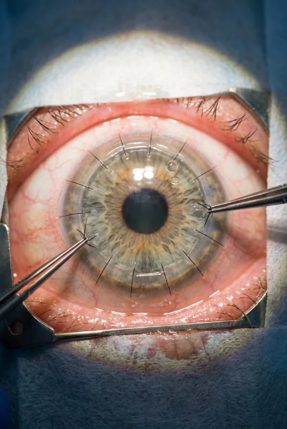
Corneal erosion (noun): corneal erosion is a break in the thin surface layer of the cornea, the epithelium, which has not stuck down firmly to the tissue beneath it. When it keeps happening it is called recurrent corneal erosion: the surface layer repeatedly loosens, typically during the night or on waking, causing sudden pain, watering and blurred vision. Most cases follow an earlier scratch to the eye or an inherited weakness of the surface layer, and a cornea specialist such as Doç. Dr. Muhammet Derda Özer can diagnose it with a short slit-lamp examination.
Corneal erosion: key facts
- What it is
- The surface layer of the cornea (epithelium) does not stay attached to its base layer, so it breaks down or peels away
- Typical warning sign
- Sharp pain, a gritty or foreign-body feeling, watering and light sensitivity, often on waking
- Most common cause
- A previous injury to the cornea: 45–64% of recurrent cases are linked to earlier trauma (EyeWiki, AAO)
- Second most common cause
- Corneal dystrophy, mainly epithelial basement membrane dystrophy (EBMD): 19–29% of cases
- Diagnosis
- Slit-lamp examination with fluorescein dye
- First-line treatment
- Preservative-free lubricating drops by day and a lubricating or hypertonic (5% sodium chloride) ointment at night
- If simple treatment fails
- Bandage contact lens, anterior stromal puncture, superficial keratectomy or phototherapeutic keratectomy (PTK)
- ICD-10-CM code (US)
- H18.831 right eye · H18.832 left eye · H18.833 both eyes · H18.839 unspecified eye
- Specialist
- Cornea specialist (ophthalmologist); at Laser Istanbul Clinic, Doç. Dr. Muhammet Derda Özer
What is corneal erosion?
The cornea is the clear front window of the eye, and its outermost layer is the epithelium, a skin of cells only a few cells thick. In a healthy eye the epithelium is anchored to the layer below it (the basement membrane and Bowman's layer) by a network of tiny attachments. In corneal erosion those attachments are weak or damaged. The epithelium can then lift, tear or slide off, leaving a raw area that exposes the cornea's very dense nerve endings. The American Academy of Ophthalmology notes that the cornea has hundreds of times more pain receptors than skin, which is why even a small erosion hurts so much.
Doctors describe two patterns. Microform erosions are milder, shorter and more frequent; macroform erosions last for days and typically follow an injury. Either way the typical story is the same: the eye feels fine, you sleep with your eyelids closed, and on waking, as the eyelid peels away from a surface that has stuck to it overnight, the loose epithelium tears. EyeWiki also notes that the lower half of the cornea is the most often affected.
Corneal erosion vs corneal abrasion: what is the difference?
An abrasion is the injury: a scratch or scrape on the surface of the cornea, for example from a fingernail, make-up brush, tree branch or eye rubbing. An erosion is what can follow: the repaired surface does not bond properly and breaks down again. When it happens two or more times it is called recurrent corneal erosion.
| Corneal abrasion | Corneal erosion (recurrent) | |
|---|---|---|
| What it is | A one-off scratch on the surface | The surface layer repeatedly loosens or tears from its base |
| Trigger | A clear injury (fingernail, branch, make-up brush, foreign body) | Often none: it can start spontaneously, most often on waking, sometimes long after an old injury |
| Typical timeline | Small abrasions heal in 1–2 days; larger ones in about a week (AAO) | Episodes come and go; the underlying weak attachment can persist for months or years without treatment |
| Treatment | Drops or ointment, sometimes an antibiotic, and follow-up | Long-term lubrication first; procedures if episodes keep returning |
| Link | A corneal abrasion is the most common starting point for recurrent erosion | May follow an abrasion, or happen in a cornea with a dystrophy |
Corneal erosion causes: why does it happen?
The most common causes and risk factors are:
Previous corneal injury
Trauma is the most common cause, accounting for 45–64% of reported recurrent cases. The injury is often something that scraped the surface, such as a fingernail, paper edge, tree branch or foreign body.
Corneal dystrophy
Inherited conditions of the cornea are the second most common cause. Epithelial basement membrane dystrophy (EBMD, also called map-dot-fingerprint dystrophy) accounts for 19–29% of cases, and some other dystrophies, such as lattice, granular or Fuchs' endothelial dystrophy, also play a part. EBMD can affect both eyes, and erosions can come on without any injury.
Dry eye and eyelid conditions
Dry eye, meibomian gland dysfunction (blocked oil glands in the eyelids), ocular rosacea and eyelids that do not close fully during sleep (nocturnal lagophthalmos) all make the surface more fragile.
Other factors
Previous corneal infection, diabetes, band keratopathy (calcium deposits on the cornea) and badly fitted or poorly maintained contact lenses are also linked to erosion.
Because the surface layer takes time to bond firmly to the layer beneath, a cornea can look healed after an abrasion and still be vulnerable for some time afterwards.
Corneal erosion symptoms: what does it feel like?
The typical corneal erosion symptoms are:
| Symptom | Notes |
|---|---|
| Sudden pain, often when you wake up or open your eyes | The classic sign. Pain can range from mild to severe |
| A feeling that something is in your eye | Gritty, scratchy, foreign-body sensation |
| Watering (tearing) | Reflex tearing as the nerves are exposed |
| Sensitivity to light (photophobia) | Many people cannot comfortably keep the eye open in bright light |
| Blurred vision | Because the surface is uneven; it usually improves as the surface heals |
| Redness | Common during an episode |
Episodes often settle within hours to days and then return after a gap that may be weeks or months. Symptoms are usually in one eye, though EBMD can affect both. The pattern of pain on waking, in someone who once scratched their eye or has a known dystrophy, is the clue that sends most people to an eye doctor.
How is corneal erosion diagnosed?
Diagnosis is usually clinical and takes a few minutes:
- History. Your doctor will ask about earlier injuries, contact lens wear, dry eye and whether pain comes on waking.
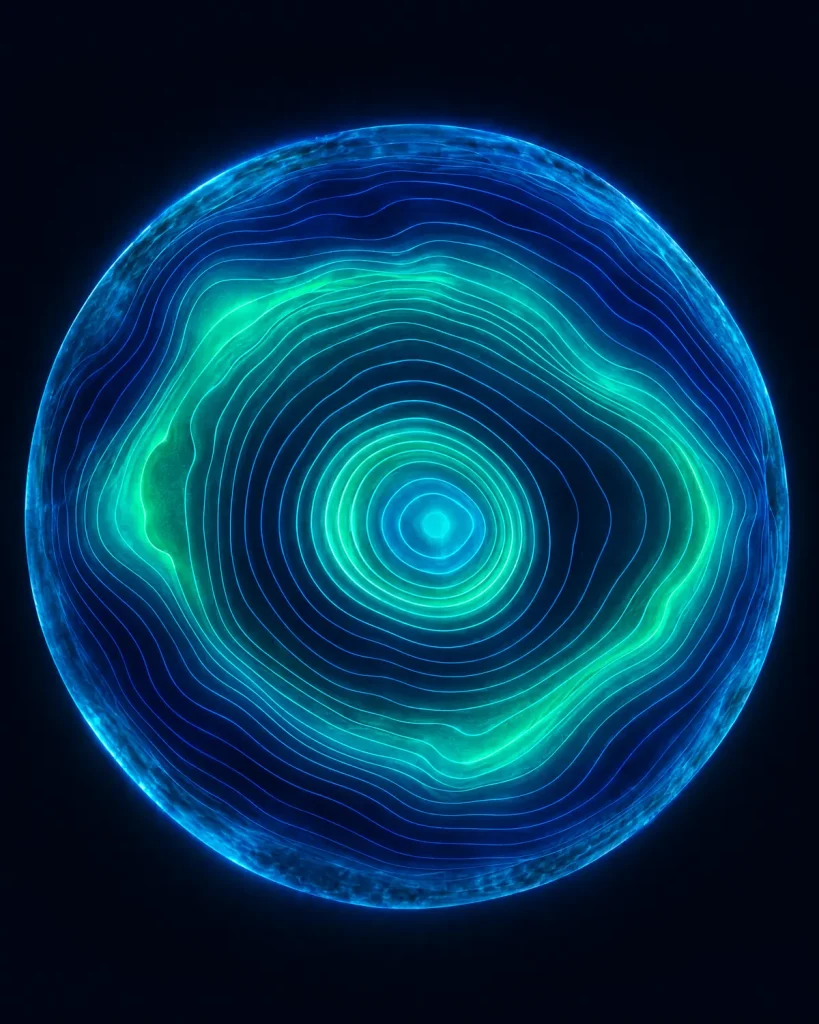
- Slit-lamp examination with fluorescein dye. The yellow-green dye shows where the surface is missing or loose. Findings range from a visible defect to a surface that looks almost normal between episodes, so the history matters.
- Corneal imaging. Optical coherence tomography (OCT) of the front of the eye can show an irregular, swollen or loosely attached epithelium. In one study of eyes with recurrent erosion, OCT showed swelling of the epithelium in about 61% and irregular break-up of the epithelium in 50% (EyeWiki).
- Checking the other eye. Dystrophies tend to show in both eyes, so the other eye is examined too.
At Laser Istanbul Clinic, corneal topography and OCT are part of every pre-operative examination in Istanbul, and your surgeon reviews your existing scans before you book.
What is the ICD-10 code for corneal erosion?
In ICD-10-CM, the clinical modification used in the United States, recurrent erosion of the cornea is coded H18.83-: H18.831 (right eye), H18.832 (left eye), H18.833 (both eyes) and H18.839 (unspecified eye). A one-off corneal abrasion is coded differently, under S05.0- (injury of conjunctiva and corneal abrasion without foreign body). Other countries use their own versions of the classification, so ask your doctor which code appears in your own records.
Corneal erosion treatment: from drops to surgery
Treatment is stepped. Most people are managed with the first steps; procedures are kept for erosions that keep returning. The 2018 Cochrane review of recurrent corneal erosion (10 trials, 505 participants) concluded that the studies were too small and of too low quality to support firm management guidelines, so your ophthalmologist will choose from the options below according to your cornea and your history.
Step 1: treating an acute episode
- Lubrication. Preservative-free artificial tears frequently during the day, and a lubricating ointment (such as a paraffin-based or hypertonic ointment) before bed. Sodium chloride 5% ointment is one of the options the American Academy of Ophthalmology names for erosions that have occurred two or more times.
- Comfort and protection. Short-term cycloplegic (pupil-relaxing) drops to ease pain, an antibiotic to prevent infection, and oral ibuprofen for discomfort.
- Don't rub. Avoid rubbing the eye, and stop wearing regular contact lenses until your ophthalmologist says it is safe (AAO).
Step 2: when erosions keep coming back (recurrent corneal erosion treatment)
| Option | What it involves | What the evidence says |
|---|---|---|
| Bandage contact lens | A soft therapeutic lens worn for weeks, with antibiotic cover, so that the surface can bond firmly | In one report, 75% of people treated for three months with an extended-wear bandage lens were free of recurrence a year later (EyeWiki). A small UK trial found no clear difference between a bandage lens and lubrication alone (Cochrane 2018) |
| Autologous serum or cyclosporine drops | Eye drops made from your own blood serum, or an anti-inflammatory drop | Second-line options (EyeWiki) |
| Oral doxycycline and short-course steroid drops | Doxycycline 50 mg twice daily blocks an enzyme (MMP-9) that breaks down the surface attachments; steroid drops are typically used two or three times a day for about 3 weeks | Used under supervision in clinical practice (EyeWiki); the quality of trial evidence is limited |
| Punctal plugs | Tiny plugs in the tear drainage openings keep drops on the eye | Help retain drops on the surface (EyeWiki) |
Step 3: corneal erosion surgery and procedures
These are options when drops, ointment and a bandage lens have not worked. Each treats the surface of the cornea; none is a corneal transplant.
| Procedure | How it works | Reported results |
|---|---|---|
| Anterior stromal puncture (micropuncture) | A fine needle (or a laser) makes tiny punctures in the loose area to help the surface bond | 62.9% success after a single treatment (EyeWiki) |
| Epithelial debridement and diamond burr polishing (superficial keratectomy) | The loose surface is removed and the layer beneath is polished so that new cells attach well | In a 48-person trial, recurrences were much less common than with a sham burr (risk ratio 0.07; moderate-certainty evidence, Cochrane 2018) |
| Alcohol delamination | Dilute alcohol (20%) loosens the surface cells for about 40 seconds, and they are wiped off | A recognised option for recurrent erosion (EyeWiki) |
| Phototherapeutic keratectomy (PTK) | An excimer laser removes a very thin layer from the front of the cornea | A 2023 meta-analysis of 700 eyes found a recurrence rate of 18%; side effects included subepithelial haze (13%), a shift towards long-sightedness (20%) and a drop in best-corrected vision (11%) |
Why surgery stays a "last step". PTK and other surface procedures can cause corneal haze or scarring, a hyperopic shift or, rarely, infection, so specialists use them only after simpler treatment has not worked. Laser treatment of the cornea also needs a healthy, thick enough cornea, which is checked with topography and pachymetry first.
Treatment with us: cornea care, including keratoconus, cross-linking and corneal transplants, is led by Doç. Dr. Muhammet Derda Özer (Doç. means Associate Professor). Corneal erosion rarely needs a transplant. If your cornea is also scarred, thinned or diseased in its deeper layers, however, a transplant may be discussed.
Corneal erosion healing time: how long does it take?
There is no single healing time, because the answer depends on whether you mean one episode or the underlying problem.
- One episode. The surface usually regrows quickly with treatment. As a guide, the AAO gives 1–2 days for a small corneal abrasion and about a week for a larger one. Pain is often worst in the first hours.
- The weak attachment. The layers need longer to bond firmly, which is why treatment continues after the pain has gone. In the studies above, bandage lens treatment lasted three months, and night-time ointment is often continued for as long as the ophthalmologist advises.
- After a procedure. After diamond burr polishing, a bandage contact lens is worn for about 4–5 days (EyeWiki); for PTK and other procedures your surgeon will give you a schedule.
Can recurrent corneal erosion be cured?
For most people it can be brought under control. EyeWiki states that with adequate treatment and prompt diagnosis, the prognosis is excellent. But recurrence is possible even after surgery (18% after PTK in the meta-analysis above), and when the cause is epithelial basement membrane dystrophy the course is variable: symptoms tend to wax and wane across a lifetime, and some people improve with time. Night-time lubrication is often recommended long term.
Does diet help recurrent corneal erosion?
No diet or food has been proven to prevent or treat recurrent corneal erosion; the EyeWiki review does not mention diet or supplements. Diet is sometimes raised because dry eye makes the surface more fragile, but the best trial on this question looked at dry eye, not erosion: in the US DREAM trial (535 people with moderate to severe dry eye), 3,000 mg of daily omega-3 fatty acids for 12 months worked no better than an olive-oil placebo. So spend your effort on what has support: lubrication, treating dry eye and eyelid disease, and not rubbing the eyes.

When to see a doctor about corneal erosion
Seek same-day care from an eye doctor or eye emergency service if you have:
- sudden severe eye pain, especially on waking, that does not ease within a few hours
- pain with a white spot on the cornea, discharge or marked redness: these can be signs of a corneal ulcer (infection), which can permanently damage vision and even cause blindness if it is not treated (AAO)
- a red, painful eye while wearing contact lenses (take the lenses out and don't put them back in)
- a chemical splash, or an injury from something sharp or fast-moving
- a sudden drop in vision
Book a routine appointment if you notice:
- repeated episodes of eye pain on waking, particularly after an old scratch
- a gritty feeling and blurred vision that return again and again
- a known corneal dystrophy such as EBMD and new episodes of pain
If you have already been treated and the episodes keep coming back, you can send your scans and eye report for a second opinion before you travel.
Frequently asked questions
Corneal erosion is a break in the surface layer of the cornea (the epithelium) that has not attached firmly to the layer beneath it. When this happens repeatedly it is called recurrent corneal erosion. It causes sudden pain, watering, light sensitivity and blurred vision, often on waking.
The main symptoms are sudden pain (often when you open your eyes after sleep), a feeling that something is in the eye, watering, sensitivity to light, redness and blurred vision. Episodes often settle within hours to days and then return.
Previous injury to the cornea is the most common cause, linked to 45–64% of recurrent cases. Corneal dystrophies, particularly epithelial basement membrane dystrophy, are the second most common cause (19–29%). Dry eye, eyelid disease and diabetes also play a part.
An abrasion is a scratch on the surface of the cornea, usually from an injury. An erosion happens when the surface layer repeatedly loosens from the cornea beneath it, often after an old abrasion or because of a dystrophy. The AAO describes erosions that have occurred two or more times as recurrent.
Treatment starts with preservative-free lubricating drops by day and a lubricating or 5% sodium chloride ointment at night. If episodes continue, options include a bandage contact lens, doxycycline with steroid drops, anterior stromal puncture, diamond burr polishing or laser treatment of the surface (PTK). Your ophthalmologist chooses the order.
Not for most people. Surgery is kept for erosions that return despite lubrication and other treatment. Options such as stromal puncture, superficial keratectomy and PTK work on the corneal surface only; they are not corneal transplants. In a meta-analysis of PTK, 18% of eyes had a recurrence.
A single episode of surface damage usually heals within days (1–2 days for a small abrasion, about a week for a larger one, according to the AAO), but the surface needs longer to bond firmly. Treatment and night-time lubrication therefore usually continue after the pain has gone.
In most people it can be controlled well, but recurrence is possible, especially if you have a dystrophy such as EBMD. Your doctor may recommend continuing night-time lubrication long term.
No diet or supplement has been shown to prevent or treat it. A large US trial found that omega-3 supplements were no better than placebo for dry eye, a common contributing factor. Lubrication, treating dry eye and avoiding eye rubbing are the measures with the best support.
In ICD-10-CM (the US version), recurrent erosion of the cornea is H18.83-, with H18.831 for the right eye, H18.832 for the left, H18.833 for both and H18.839 for an unspecified eye. A single corneal abrasion is coded under S05.0-.
Sources for the facts on this page
- EyeWiki (American Academy of Ophthalmology). Recurrent Corneal Erosion. https://eyewiki.org/Recurrent_Corneal_Erosion
- American Academy of Ophthalmology. Corneal Abrasion and Erosion. https://www.aao.org/eye-health/diseases/what-is-corneal-abrasion
- American Academy of Ophthalmology. What Is a Corneal Ulcer (Keratitis)? https://www.aao.org/eye-health/diseases/corneal-ulcer
- EyeWiki (American Academy of Ophthalmology). Epithelial Basement Membrane Dystrophy. https://eyewiki.org/Epithelial_Basement_Membrane_Dystrophy
- Watson SL, Leung V. Interventions for recurrent corneal erosions. Cochrane Database of Systematic Reviews 2018, CD001861.pub4. PubMed: https://pubmed.ncbi.nlm.nih.gov/29985545/
- Chen S, Chu X, Zhang C, et al. Safety and Efficacy of the Phototherapeutic Keratectomy for Treatment of Recurrent Corneal Erosions: A Systematic Review and Meta-Analysis. Ophthalmic Research 2023;66(1):1114–1127. PubMed: https://pubmed.ncbi.nlm.nih.gov/37490883/
- Asbell PA, Maguire MG, et al. (DREAM Study Research Group). n-3 Fatty Acid Supplementation for the Treatment of Dry Eye Disease. New England Journal of Medicine 2018;378:1681–1690. PubMed: https://pubmed.ncbi.nlm.nih.gov/29652551/
- Ewald M, Hammersmith KM. Review of diagnosis and management of recurrent erosion syndrome. Current Opinion in Ophthalmology 2009;20(4):287–291. PubMed: https://pubmed.ncbi.nlm.nih.gov/19491682/
- US National Library of Medicine, Clinical Tables. ICD-10-CM search service (H18.83-, S05.0-). https://clinicaltables.nlm.nih.gov/apidoc/icd10cm/v3/doc.html
Repeated eye pain on waking, or a cornea problem that keeps returning?
Send us your scans or eye report. Doç. Dr. Muhammet Derda Özer, our cornea specialist, will review them and tell you which treatments may suit your eyes, with no obligation.
3 eye surgeons
Your case is reviewed by a named surgeon, not a salesperson. Reply within {{response time}}.
 Prof. Dr. Ahmet Satıcı
 Doç. Dr. Muhammet Derda Özer
 Op. Dr. Aysan Bayram