
- Home
- Knowledge centre
- Retina & glaucoma guides
- Sudden Floaters in One Eye: When to Worry and When Vitrectomy Helps
Sudden Floaters in One Eye: When to Worry and When Vitrectomy Helps
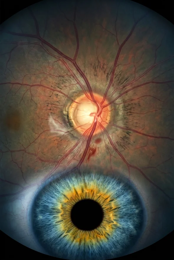
Sudden floaters in one eye need an eye examination the same day, because they can mean a retinal tear or detachment. Most are caused by the vitreous gel separating from the retina (posterior vitreous detachment), which is harmless alone, but only a dilated examination can tell the difference. Long-standing floaters rarely need treatment; vitrectomy is for severe cases. In Istanbul, Doç. Dr. (Associate Professor) Muhammet Derda Özer or Op. Dr. Aysan Bayram assess them.
Sudden floaters in one eye: when to worry
Get an eye examination today if you notice a sudden new floater, a shower of floaters, flashes of light, or a shadow or curtain across your vision. The NHS, the American Academy of Ophthalmology (AAO) and the US National Eye Institute (NEI) all give this advice.
The NHS tells you to contact an optician urgently or call NHS 111 for any of these:
- floaters or flashes that appear for the first time
- a sudden increase in the number of floaters or flashes
- a dark curtain or shadow moving across your vision
- blurred vision or eye pain along with floaters or flashes
- floaters after eye surgery or an eye injury
The AAO adds a dark shadow or blurry area in your side or central vision. Outside the UK, go to an ophthalmologist or an emergency eye service the same day. Do not delay treatment in order to travel. If you are already in Turkey, message us and we will arrange an urgent retina examination {{urgent appointment availability}}.
The reason for the urgency is simple. The vitreous, the clear gel inside your eye, can tug on the retina as it shrinks. If the pull tears the retina, fluid can pass through the tear and lift the retina off the back of the eye. That is a retinal detachment, which can cause permanent loss of vision if it is not treated. A tear found early can be sealed with a laser; a detachment needs an operation.
What causes sudden floaters in one eye?
The most common cause of sudden floaters in one eye is posterior vitreous detachment (PVD), a normal age-related change in which the vitreous gel pulls away from the retina. The NHS names PVD as the main cause of floaters and flashes. The AAO says it usually happens to most people by the age of 70, and NHS eye-clinic guidance says it typically happens after the age of 40. As the gel becomes more liquid and shrinks, collagen strands clump together and cast shadows on the retina. Those shadows are the floaters you see.
Other causes are less common but more serious, which is why a sudden floater always needs an examination:
| Cause | What happens | Why it matters |
|---|---|---|
| Posterior vitreous detachment (PVD) | The vitreous separates from the retina and may leave a floating ring or strands | Usually harmless; can tear the retina as it separates |
| Retinal tear | The vitreous pulls a hole in the retina | Can progress to retinal detachment if not sealed |
| Retinal detachment | The retina peels away from the back of the eye | Emergency; can cause permanent sight loss |
| Bleeding into the vitreous | Blood from a torn retinal vessel, diabetes or injury shows up as dark floaters | Often points to a tear or to diabetic eye disease |
| Inflammation (uveitis) or infection | Inflammatory cells float in the vitreous | The underlying cause needs treatment |
| Eye injury or previous eye surgery | Disturbs the vitreous or retina | The NHS lists floaters after surgery or injury as urgent |
Some people are more likely to be affected. The NEI names people who are very nearsighted, have diabetes or have had cataract surgery. EyeWiki (the AAO's clinical encyclopedia) adds that PVD comes earlier in nearsighted eyes, with inflammation, after blunt trauma and after cataract surgery. Floaters are also very common: in a 2013 survey of 603 smartphone users, 76% reported seeing floaters and 33% said they noticeably affected their vision.
How often does a sudden floater mean a retinal tear?
Not every sudden floater is dangerous, but a meaningful minority are. In a 2022 study of 8,305 adults with new symptoms of PVD in a US health system, 5.4% had a retinal tear and 4.0% had a retinal detachment. In a retina-specialist practice (7,999 eyes, 2020 study), 16.0% had a retinal break and 6.2% a detachment at the first examination. A 2017 study of 365 people with PVD symptoms found a retinal tear in 14.5% and bleeding in the vitreous or retina in 22.7%.
In these studies, a higher risk went with blurred vision, vitreous haemorrhage and lattice degeneration (a thinning of the peripheral retina). Flashes did not raise the risk. In other words, you cannot judge your own risk from how a floater looks, which is why the examination matters.

What is a sudden large floater in one eye?
A single large floater that appears suddenly is often a Weiss ring, but it can also be a sign of bleeding or a retinal tear, so it needs the same-day check. A Weiss ring is a ring-shaped floater. EyeWiki describes it as forming at the edge of the vitreous where it separates from around the optic nerve. Like other floaters, it drifts through your vision and shows up most against a plain bright background such as the sky or a white wall.
A large floater that comes with flashes, many smaller floaters or a shadow is more worrying than a large floater alone. The examination looks at the whole retina, not only at what you can see.
Sudden floaters in one eye and headache: is there a link?
A headache with sudden floaters does not make the floaters safe or dangerous, so judge the eye symptom, not the headache. A new floater or flashes need an examination even if you also have a headache, and the NHS includes eye pain with floaters on its urgent list.
The AAO describes one harmless cause of flashes: migraine. Migraine flashes look like jagged lines, shimmering spots or heat waves, can last up to 60 minutes and can happen without any headache ("ocular migraine"). Flashes caused by the vitreous rubbing or pulling on the retina are different, and floaters are dark shapes that drift as your eye moves (NEI).
Seek help the same day if:
- you have a new floater or new flashes, with or without a headache
- the headache comes with eye pain, redness or blurred vision
- you notice a shadow, a curtain or any sudden loss of vision
What are the 4 types of eye floaters?
There is no official list of four types, but people most often describe floaters as dots or spots, threads or strings, cobwebs and rings. The AAO says floaters look like "small specks, dots, circles, lines or cobwebs", and the NEI describes spots, threads, squiggly lines and cobwebs. EyeWiki describes the ring shape as a Weiss ring.
| Shape | What it looks like | Usual cause |
|---|---|---|
| Dots or spots | Small dark specks that drift | Small clumps of vitreous gel or cells |
| Threads or strings | Squiggly lines or short strands | Collagen fibres that have clumped together |
| Cobwebs | Fine, web-like shadows | Fibres in the shrinking gel; sometimes blood or cells |
| Rings (Weiss ring) | A ring or oval, often large | Left where the vitreous separated from the optic nerve area |
The shape alone does not tell you whether a floater is safe. A sudden shower of tiny dots is a classic warning sign, while a ring that appears suddenly is often a PVD. Both need a dilated examination.
A single black spot in your vision: is it a floater?
If the spot stays in one place in your vision instead of drifting, it may not be a floater. Floaters move as your eye moves, and the NEI says that when you try to look straight at them, they appear to move away. They are most noticeable against bright, plain backgrounds (AAO).
A dark spot that stays fixed, or that sits in the centre of your vision, is a different problem. For example, the NEI describes how a macular hole, a break in the central retina, starts as blurry or wavy vision and can progress to a blind spot in the central vision, and says treatment works best when it is found early. Several retinal conditions can cause a fixed dark or blurred area, and only an examination can tell which one it is.
| A floater | A fixed black spot | |
|---|---|---|
| Movement | Drifts and moves away when you look at it | Stays in the same place in your vision |
| Best seen against | A bright, plain surface | Not tied to the background |
| Other signs | May come with flashes | May come with blurred or wavy vision |
| What to do | New or sudden: examination today | Examination as soon as possible |
What happens at the eye examination?
The doctor widens your pupil with drops and examines the whole retina. The NEI calls this a dilated eye examination. EyeWiki describes the standard method as indirect ophthalmoscopy with scleral indentation, plus a slit-lamp examination, so the doctor can see the far edge of the retina where tears often start.
The examination usually finds one of three things:
- A PVD with no tear. No treatment is needed, and you are told which warning signs to watch for. EyeWiki describes a first follow-up about one month after diagnosis, then checks every 3 to 6 weeks for 3 months.
- A retinal tear. The tear is sealed with a laser or freezing treatment (laser retinopexy or cryotherapy) so it does not become a detachment.
- A retinal detachment or heavy bleeding. You need surgery, usually vitrectomy, which removes the vitreous and repairs the retina from inside the eye.
A normal first examination is reassuring but not the end of the story. In a 2020 study of 7,999 eyes with acute PVD, a new retinal break appeared later in 2.6% and a retinal detachment in 1.0%, and almost half of the delayed breaks and two-thirds of the delayed detachments were found more than six weeks after the first visit. A 2022 study found that late tears or detachments occurred in 12.4% of people who had vitreous haemorrhage, lattice degeneration or a tear or detachment in the other eye at the start, but only 0.7% of those without any of these. This is why you are told to return at once if your symptoms change, even after a normal check.
Sudden floaters in one eye: treatment
Treatment depends on the cause, not on the floater. The NHS says you will usually only need treatment if you have a problem that could affect your vision.
| Cause found | Treatment |
|---|---|
| PVD only, no tear | Observation and warning-sign advice; no medical therapy is recommended (EyeWiki) |
| Retinal tear | Laser retinopexy or cryotherapy to seal the tear |
| Retinal detachment | Surgery: vitrectomy, scleral buckle or gas injection, chosen by the surgeon |
| Bleeding in the vitreous | Treat the cause (a tear, diabetic eye disease); vitrectomy if the blood does not clear |
| Inflammation (uveitis) | Treatment of the inflammation or infection |
| Long-standing floaters that bother you | Adapting first; vitrectomy or laser only in selected severe cases (see below) |
Do floaters go away, and can you reduce them naturally?
Most floaters become less noticeable over time, but they do not usually disappear. The NHS says floaters often become less noticeable as you adjust to them, and flashes may eventually stop. The AAO says they typically fade and become less noticeable, and the NEI says that if floaters come with ageing and do not bother you, you probably will not need treatment.
How to reduce floaters in your eyes naturally
No natural remedy has been shown to remove floaters, so for most people the honest answer is to adapt to them. None of the NHS, AAO or NEI pages we reviewed lists a home remedy, eye exercise or eye drop as a way to clear floaters. What does help:
- Have the first episode checked. Once a doctor has confirmed that the retina is intact, most people notice floaters less.
- Move your eyes. When a floater drifts into your line of sight, look up and down or side to side; floaters move away when you look straight at them (NEI).
- Reduce bright, plain backgrounds. Floaters show most against bright, plain surfaces (AAO). Dimming a screen or wearing sunglasses outdoors may make them less visible; this is a practical tip, not a treatment.
- Know your warning signs. Watching for a sudden change in your floaters is the best protection for your sight.
Do eye health supplements help floaters?
There is no supplement for floaters recommended in the guidance we reviewed. The NHS, AAO and NEI do not recommend vitamins or supplements as a treatment for floaters, and EyeWiki says no medical therapies are recommended for PVD. Be cautious about products that promise to dissolve floaters. Supplements are sometimes discussed for other eye conditions, but that is a different question. Ask your eye doctor before buying a product marketed for floaters.

Laser eye surgery for floaters: does YAG vitreolysis work?
YAG laser vitreolysis uses a laser to break up floaters inside the eye, and the evidence is promising but limited. In a masked, sham-controlled trial of 52 patients with floaters from a Weiss ring (JAMA Ophthalmology, 2017), 54% of those treated reported improved symptoms, against 9% of the sham group, and no retinal tears or detachments occurred in the treated group. The trial was small, followed patients for only 6 months and could not rule out less common complications. People with a history of retinal tear or detachment, uveitis, diabetic retinopathy or glaucoma were excluded.
Case reports also describe rapid cataract progression and glaucoma that was hard to control after floater laser. A 2016 review in Survey of Ophthalmology found that reports of resolution with laser ranged from 0% to 100%, and concluded that at that time only vitrectomy had proven value.
The practical points:
- The trial studied Weiss ring floaters, so its results may not apply to other types.
- Laser is not a treatment for sudden floaters, tears or detachment.
- Ask any clinic offering it who performs it, how many they have done and what happens if the result is poor. Whether our surgeons offer it: {{YAG laser vitreolysis availability}}.
When does vitrectomy help eye floaters?
Vitrectomy can clear severe floaters that have lasted for months and interfere with daily life, but it is seldom necessary and it has real risks. The AAO states that severe floaters can be removed by surgery, "but this has risks and is seldom necessary or recommended". The NEI says to talk with your doctor about the risks and benefits if floaters make it hard to see clearly and interfere with daily life.
In a vitrectomy, the surgeon removes the vitreous gel through tiny openings in the white of the eye, and the floaters go with it. For floaters, this is often a limited, central removal rather than the whole gel. The gel is replaced with clear saline solution. It is usually done under local anaesthetic.
What do the studies show?
A 2022 systematic review of 18 studies (2,077 eyes) found that at least 90% of patients were satisfied or had relief of symptoms. The largest multicentre study in it found 92% were satisfied or extremely satisfied, 6% neutral and 2% dissatisfied. A survey of 581 eyes operated on by 48 surgeons in 16 countries (European VitreoRetinal Society, EVRS) found 92.4% satisfied or extremely satisfied. The same survey found that patients whose floaters were small or almost invisible before surgery were less satisfied.
What are the risks of vitrectomy for floaters?
| Complication | Pooled rate in the 2022 meta-analysis | EVRS survey (581 eyes) |
|---|---|---|
| Cataract | 31.7% | 48.6% of eyes with their own lens needed cataract surgery, at a median of 16 months |
| Retinal tear or break | 2.9% | 5% (17% with 20-gauge, 5.4% with 23-gauge instruments) |
| Retinal detachment | 1.5% | 2.4%, at a median of 3 months |
| Bleeding into the vitreous | 2.0% | not reported |
| Macular swelling | 1.7% | 1.4% (cystoid macular oedema) |
| Infection inside the eye (endophthalmitis) | 0.18% | none reported |
Figures are from published studies (see Sources); your own risks depend on your eye. The studies followed patients for different lengths of time.
What this means for you: the biggest practical trade-off is cataract. If you still have your natural lens, there is a real chance you will need cataract surgery within a couple of years of the operation. The surgeon also weighs the small but serious risk of a retinal tear or detachment. That is why vitrectomy is normally recommended only when floaters clearly affect your quality of life, not for occasional annoyance.
Who is a candidate?
Vitrectomy for floaters is usually considered when:
- floaters have been present for months and have not become easier to ignore
- they interfere with reading, driving, work or other daily activities
- a retina specialist has examined the retina and found it healthy
- you understand the risks, including cataract, and accept them
It is not suitable for new, sudden floaters (these need a diagnosis first), nor for floaters that are small, rare or barely visible.

Eye floaters treatment in Istanbul
If you have long-standing floaters that spoil your vision, Doç. Dr. Muhammet Derda Özer or Op. Dr. Aysan Bayram can assess your eyes and tell you honestly whether surgery is worth it. Both work with retinal disease and vitrectomy. You send your scans and history through the free online consultation, the surgeon reviews them, and you get an answer before you book. Sometimes that answer is "your retina is healthy and surgery is not needed".
Surgery takes place in a fully equipped, Ministry of Health–licensed operating theatre in Istanbul. Vitrectomy with Laser Istanbul Clinic costs from {{£ price}} per eye. The price includes the pre-operative examination and scans, surgery by your named surgeon, medication, follow-up checks in Istanbul, a personal interpreter, a coordinator and remote follow-up. Hotel and airport transfers are not included; we arrange them on request at a separate charge. Flights are not included.
If your symptoms are new, do not travel for them: be examined first where you are. Once your retina has been checked, we are glad to review your scans.
Frequently asked questions
They can be. A sudden new floater, a shower of floaters, flashes or a curtain across your vision can mean a retinal tear or detachment, and the NHS, AAO and NEI all advise an urgent examination. Most turn out to be a harmless PVD, but you cannot tell without a dilated examination. Go the same day.
Most often a posterior vitreous detachment, where the vitreous gel separates from the retina with age. Less often it is a retinal tear or detachment, bleeding into the vitreous, inflammation (uveitis), infection, or the effect of an eye injury or previous eye surgery.
Yes, but the two may be unrelated. Migraine can cause flashes, described by the AAO as jagged lines or shimmering spots lasting up to an hour, sometimes without a headache. A new floater, new flashes or eye pain still need an examination, whatever the headache.
There is no fixed time. The NHS says floaters often become less noticeable over time as you adjust to them, and flashes may eventually stop. Some floaters remain, but most people stop noticing them. Any sudden change should be checked again.
Sometimes. In a 2017 randomised trial of 52 patients, 54% improved after YAG laser against 9% after sham treatment. The evidence is limited, and laser is not suitable for every floater. Ask your retina specialist whether your floater is a good target.
It is generally effective but not risk-free. In a 2022 meta-analysis, retinal detachment occurred in about 1.5% of eyes, retinal tears in about 2.9% and infection in about 0.18%. Cataract is the most common problem: about 32% pooled, and 48.6% of eyes with their own lens in a European survey.
No natural remedy or supplement has been shown to remove floaters, and the NHS, AAO and NEI do not recommend one. Adapting, moving your eyes and having a doctor confirm that your retina is healthy are what help most people.
People usually describe dots or spots, threads or strings, cobwebs and rings (Weiss ring). This describes shapes, not an official classification, and the shape does not show whether a floater is dangerous.
Yes, if a retina examination shows that your floaters are severe and your retina is healthy. Doç. Dr. Özer or Op. Dr. Bayram review your scans first. For sudden floaters, flashes or a curtain, have a local emergency eye examination first.
Floaters that spoil your vision? Get a retina surgeon's opinion before you decide.
Our retina surgeons, Doç. Dr. Muhammet Derda Özer and Op. Dr. Aysan Bayram, review your scans, explain whether your retina is healthy and tell you plainly whether vitrectomy is worth the risk. You get a written plan and a price in writing before you travel.
Sources for the facts on this page
- NHS. Floaters and flashes in the eyes. https://www.nhs.uk/symptoms/floaters-and-flashes-in-the-eyes/
- American Academy of Ophthalmology. What Are Floaters and Flashes? https://www.aao.org/eye-health/diseases/what-are-floaters-flashes
- American Academy of Ophthalmology. What Is Posterior Vitreous Detachment? https://www.aao.org/eye-health/diseases/what-is-posterior-vitreous-detachment
- National Eye Institute (NIH). Floaters. https://www.nei.nih.gov/learn-about-eye-health/eye-conditions-and-diseases/floaters
- National Eye Institute (NIH). Macular Hole. https://www.nei.nih.gov/learn-about-eye-health/eye-conditions-and-diseases/macular-hole
- EyeWiki (AAO). Posterior Vitreous Detachment. https://eyewiki.org/Posterior_Vitreous_Detachment
- Moorfields Eye Hospital NHS Foundation Trust. Flashes and floaters. https://www.moorfields.nhs.uk/condition/flashes-and-floaters
- Complications of Acute Posterior Vitreous Detachment (8,305 patients, Kaiser Permanente Northern California). Ophthalmology 2022. PMID 34324945. https://pubmed.ncbi.nlm.nih.gov/34324945/
- Delayed Retinal Breaks and Detachments after Acute Posterior Vitreous Detachment (7,999 eyes). Ophthalmology 2020. PMID 31767432. https://pubmed.ncbi.nlm.nih.gov/31767432/
- Posterior vitreous detachment: prevalence of and risk factors for retinal tears. Clinical Ophthalmology 2017. https://pmc.ncbi.nlm.nih.gov/articles/PMC5609787/
- Efficacy and Safety of Pars Plana Vitrectomy for Primary Symptomatic Floaters: A Systematic Review with Meta-Analyses. Ophthalmology and Therapy 2022. https://pmc.ncbi.nlm.nih.gov/articles/PMC9587141/
- Management of vitreous floaters: an international survey, the European VitreoRetinal Society Floaters study report. Eye 2020. https://pmc.ncbi.nlm.nih.gov/articles/PMC7182575/
- YAG Laser Vitreolysis vs Sham YAG Vitreolysis for Symptomatic Vitreous Floaters: A Randomized Clinical Trial. JAMA Ophthalmology 2017. https://pmc.ncbi.nlm.nih.gov/articles/PMC5710539/
- Vitreous floaters: Etiology, diagnostics, and management. Survey of Ophthalmology 2016. PMID 26679984. https://pubmed.ncbi.nlm.nih.gov/26679984/
- Refractory open-angle glaucoma after neodymium-yttrium-aluminum-garnet laser lysis of vitreous floaters. American Journal of Ophthalmology 2015. PMID 25308785. And: Rapid Cataract Progression after Nd:YAG Vitreolysis for Vitreous Floaters: A Case Report and Literature Review. Case Reports in Ophthalmology 2017. https://pmc.ncbi.nlm.nih.gov/articles/PMC5471782/
- Prevalence of vitreous floaters in a community sample of smartphone users. International Journal of Ophthalmology 2013. https://pmc.ncbi.nlm.nih.gov/articles/PMC3693028/