
- Home
- Knowledge centre
- Eye glossary A–Z
- Fovea Centralis and Macula: Location, Function and Diseases
Fovea Centralis and Macula: Location, Function and Diseases
Medically reviewed by {{surgeon}}, {{surgeon's title and specialty}}, on {{review date}}. Last updated {{date}}.
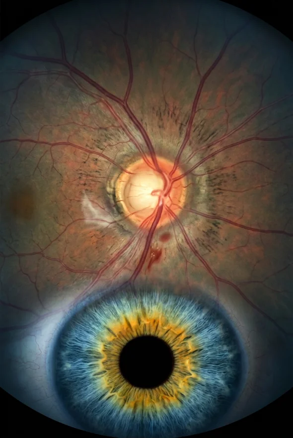
Fovea centralis (noun, plural foveae; also "fovea of the eye"): the fovea centralis is a tiny pit in the centre of the macula, the central part of the retina, where the eye sees in the sharpest detail. The macula (Latin macula lutea, "yellow spot") is the retinal area around it that controls sharp, straight-ahead vision, the vision you use to read, recognise faces and drive. Together they are the reason you can see fine print and colour wherever you point your eyes.
In short: the macula is about 5–6 mm across; the fovea centralis, about 1.5 mm wide at its centre, is its sharpest point. Damage to either blurs or distorts central vision while side vision stays. Common causes are age-related macular degeneration, macular hole, macular pucker, diabetic macular oedema and central serous retinopathy. Retina conditions are assessed in Istanbul by Doç. Dr. Muhammet Derda Özer (Doç. = Associate Professor) and Op. Dr. Aysan Bayram.
Macula and fovea: key facts
- Where they are
- At the back of the eye, in the centre of the retina, on the temple side of the optic nerve head (the optic disc)
- Macula size
- About 5–6 mm in diameter
- Fovea size
- About 1.5 mm in diameter, in the middle of the macula
- Foveola
- The very centre of the fovea, about 350 µm (0.35 mm) across and 90–130 µm thick
- Photoreceptors
- Cones only at the foveola; no rods, so no dim-light cells at the exact centre
- Blood vessels
- None at the centre: a foveal avascular zone of roughly 0.2–1 mm
- What they do
- Sharp central vision, fine detail and colour
- Typical symptoms of damage
- Blurred or distorted centre of vision, straight lines looking wavy, a grey or dark patch in the middle
- Specialists
- Retina specialist (ophthalmologist); at Laser Istanbul Clinic, Doç. Dr. Muhammet Derda Özer and Op. Dr. Aysan Bayram
What is the fovea centralis?
The fovea centralis is the small depression at the very centre of the macula, and it gives the eye its sharpest vision. It is only about 1.5 mm across, yet it matters far more than its size suggests: in a 2025 review of macular anatomy, researchers noted that approximately 25% of all retinal ganglion cells, the nerve cells that carry the picture from the eye to the brain, are located in the fovea.
The fovea has several features that set it apart from the rest of the retina:
- It is a pit. Around the centre of the fovea, the inner layers of the retina are pushed to the sides, leaving only the thinnest sheet of retina, mostly cone cells, at the very bottom. Light therefore reaches the photoreceptors with little tissue in the way.
- It is packed with cones. Cones are the photoreceptors that work in good light and see colour and detail. At the fovea they sit at their highest density, arranged in a tight hexagonal pattern, and the rods (the cells for dim light) are excluded.
- It has no blood vessels at its centre. This is the foveal avascular zone, which measures roughly 0.2 to 1 mm across in humans.
- It holds yellow pigment. The highest concentration of macular pigment (lutein, zeaxanthin and meso-zeaxanthin) is found at the fovea. These pigments give the macula its yellowish colour, hence macula lutea.
Where is the fovea located?
The fovea sits at the back of the eye, in line with the centre of your gaze. It lies on the temple side of the optic disc, the spot where the optic nerve leaves the eye: about 4.5–5 mm away, or roughly two and a half disc diameters.
Glossary: Optic nerveWhat is the foveola?
The foveola is the innermost part of the fovea: about 350 µm across and 90–130 µm thick. It is made up exclusively of cone photoreceptors and contains no rods, no ganglion cells and no short-wavelength-sensitive (blue) cones. It is where visual sharpness peaks.
Macula vs fovea: what is the difference?
The two words are often used as if they meant the same thing, but they describe nested areas of the same patch of retina, from largest to smallest:
| Term | Size | What it is | Why it matters |
|---|---|---|---|
| Macula (macula lutea) | About 5–6 mm across | The central retina, rich in cones, yellow with macular pigment | Controls sharp, straight-ahead vision: reading, faces, driving, colour |
| Fovea (fovea centralis) | About 1.5 mm across | The pit in the middle of the macula | Highest cone density; best detail vision |
| Foveola | About 0.35 mm across | The floor of the fovea | Cones only; the exact point you look at |
An easy way to remember it: the macula is the area, the fovea is the pit in its middle, and the foveola is the floor of that pit. Eye doctors speak of "macular disease" for any condition of this region, and of the fovea being involved when the very centre is affected.

What do the macula and fovea do?
The macula gives you sharp, detailed, central vision; the fovea is its sharpest point. The National Eye Institute describes the macula as the part of the eye that controls sharp, straight-ahead vision. In everyday life this covers reading, recognising faces, seeing road signs, threading a needle and seeing colour.
That is why you turn your eyes towards whatever you want to see clearly: only the small area imaged on the fovea is seen in the sharpest detail, and the rest of what you see is built from your side vision.
Side (peripheral) vision comes from the rest of the retina, which is dominated by rods. It is poorer at detail but better suited to dim light. Macular disease therefore blurs or blocks the middle of what you look at while leaving the edges of your vision. It does not cause total blindness, but it can make reading, driving and recognising faces hard.
Which diseases affect the macula and fovea?
Age-related macular degeneration (AMD)
AMD is a common condition that affects the middle part of your vision and usually first appears in people in their 50s (NHS). The National Eye Institute estimates that 11 million people in the United States have it. It comes in two forms. Dry AMD is the more common one: the macula gradually gets thinner with age, usually over several years, through early, intermediate and late stages. Wet AMD is less common but usually causes faster vision loss: abnormal blood vessels grow at the back of the eye and damage the macula. The exact cause is not known, and risk factors include ageing, smoking, high blood pressure, being overweight and a family history of AMD.
Macular degeneration treatment & Scharioth macula lensMacular hole
A macular hole is a small circular opening that forms in the macula. The leading cause is age: as the gel inside the eye (the vitreous) shrinks, it can stay stuck to the retina and stretch the macula until it opens (American Academy of Ophthalmology). It mostly affects people aged 60 and over and is more common in women. EyeWiki reports an incidence of about 8.69 eyes per 100,000 each year. Doctors grade macular holes in four stages: in stage 1 the normal dip of the fovea is lost, and in stage 3 the hole is 400 µm or wider.
Macular pucker (epiretinal membrane)
A macular pucker forms when wrinkles, creases or bulges develop on the macula, so it is no longer flat. The cause is usually age-related, from the same vitreous shrinkage as macular hole, but it can also follow eye inflammation, previous eye surgery, diabetes and other retinal problems. Vision becomes wavy, blurred or distorted, but side vision is not affected.
Diabetic macular oedema (DME)
In diabetic macular oedema, blood vessels in the retina leak fluid into the macula. About 1 in 15 people with diabetes eventually develop it (National Eye Institute). The main symptom is blurred central vision.
Central serous chorioretinopathy
In this condition fluid collects under the retina, usually in one eye. It mainly affects people aged 35–55 and is more common in men. Risk factors include corticosteroid use, sleep apnoea, high stress levels and pregnancy. Most cases clear up on their own within about three months, but around half come back, so follow-up matters.
Symptoms of macula and fovea problems
Macular disease is usually painless and does not change the look of the eye. The signs below come from the NHS, the National Eye Institute and the American Academy of Ophthalmology:
| Symptom | What it can point to |
|---|---|
| A blurred or distorted area in the centre of vision | AMD, macular hole, macular pucker, diabetic macular oedema, central serous retinopathy |
| Straight lines looking wavy or crooked | AMD, macular pucker, macular hole, central serous retinopathy |
| A dark, grey or blank spot in the middle of vision | Macular hole, advanced AMD, central serous retinopathy |
| Trouble seeing in low light, or colours looking duller | AMD, central serous retinopathy |
| Objects looking smaller or further away than they are | Central serous retinopathy |
| Difficulty reading or recognising faces while side vision stays normal | Any macular disease |
Early dry AMD often causes no symptoms at all, so it is typically found at a routine eye examination.

How are the macula and fovea examined?
An eye specialist looks at the macula with a few quick, painless tests:
- Dilated eye examination. Drops widen the pupil so the doctor can see the back of the eye, including the macula.
- Optical coherence tomography (OCT). OCT is a non-contact scan that produces a cross-section picture of the retina. Spectral-domain OCT has a resolution of less than 5 µm and shows the layers of the macula, the foveal pit, fluid, holes and puckers. Ophthalmologists use it to diagnose macular hole and macular pucker (AAO).
- Angiography. In some cases a dye test (fluorescein angiography) is used to show leaking or abnormal blood vessels, for example in central serous retinopathy.
At Laser Istanbul Clinic, OCT scanning is part of every pre-operative eye examination in Istanbul, and your surgeon will review OCT scans you already have before you book.
How are macular diseases treated?
Treatment depends on the condition, and not all damage can be reversed. In general, treatment aims to protect the sight you still have and, where possible, to improve it.
| Condition | Typical treatment | What to know |
|---|---|---|
| Wet AMD | Anti-VEGF injections into the eye; in some cases photodynamic therapy | Treatment is started quickly; injections are usually repeated over time |
| Dry AMD | Dietary supplements for intermediate AMD; monitoring | Aims to slow progression, not cure it |
| Advanced AMD | Low-vision aids and, in selected patients, a magnifying lens implant | The Scharioth macula lens is an add-on magnifier for reading, not a cure |
| Macular hole | Vitrectomy (removing the vitreous gel) with a gas bubble | The bubble holds the hole closed while it heals |
| Macular pucker | Observation if mild; vitrectomy with membrane peeling if severe | Vision may improve gradually but may not fully recover |
| Diabetic macular oedema | Anti-VEGF or corticosteroid injections, or laser | Treatment aims to stop vision getting worse; control of diabetes, blood pressure and cholesterol also matters |
| Central serous retinopathy | Usually watching and waiting; laser, photodynamic therapy or tablets if it persists | Most cases settle on their own |
Macular hole surgery: what to expect
For a macular hole, the American Academy of Ophthalmology recommends vitrectomy surgery: the surgeon removes the vitreous gel pulling on the macula and places a gas bubble inside the eye to press the hole closed while it heals. Patients may need to hold a particular head position for up to a week, and must avoid changes in altitude, including flying, until the bubble has gone. Risks include infection, bleeding, retinal detachment, raised eye pressure and cataract. Because the gas affects when you can travel, plan your dates with your surgeon first. Surgery takes place in a fully equipped, Ministry of Health–licensed operating theatre in Istanbul.
Treatment with us: macular conditions are assessed and treated by Doç. Dr. Muhammet Derda Özer (retina) and Op. Dr. Aysan Bayram (retinal disease), and both also perform cataract surgery. Your examination always includes an OCT scan before any recommendation is made.

When to see a doctor about your macula
Seek same-day care from an eye doctor or eye emergency service if:
- your vision suddenly gets worse
- you see a dark curtain or shadow moving across your vision
- your eye is red and painful
(These are NHS warning signs that can point to other eye problems needing urgent treatment.)
Book a prompt appointment if you notice:
- a blurred, distorted or missing patch in the centre of your vision
- straight lines, such as door frames or text, looking bent or wavy
- reading getting harder even with new glasses
- colours looking faded, or needing much more light to read
Check each eye separately by covering the other one. If you already have a diagnosis, such as macular degeneration, and want a second opinion on treatment, you can send your OCT scans for review before you travel.
Frequently asked questions
The fovea centralis is a small pit in the centre of the macula, about 1.5 mm across, where cone cells are packed most densely. It gives the eye its sharpest detail vision, and it is what you point at whatever you want to see clearly.
The macula is the whole central area of the retina, about 5–6 mm across, and it controls sharp, straight-ahead vision. The fovea is the pit in the middle of the macula, about 1.5 mm across, where vision is sharpest.
At the back of the eye, in the centre of the retina and in line with your gaze. It lies about 4.5–5 mm from the optic disc, on the temple side.
The fovea provides your sharpest, most detailed vision. It is made up mostly of tightly packed cone cells, which handle detail and colour in good light, and the rods are excluded from it.
Cones. The foveola, the very centre of the fovea, is exclusively populated by cones. Rods, which handle dim light, are found mainly in the outer retina.
Because of its design: a thin pit with little tissue over the photoreceptors, very densely packed cones, and a large share of the retina's nerve cells dedicated to carrying signals from this tiny area.
Central vision becomes blurred, distorted or patchy, and straight lines may look wavy. Side vision usually stays normal, so macular disease alone does not cause total blindness.
Sometimes. A macular hole or macular pucker can be repaired with surgery, and wet AMD and diabetic macular oedema can be treated with injections to protect or improve sight. Dry AMD cannot be cured, but its progression can be slowed. Your retina specialist can tell you which applies to you after an OCT scan.
AMD is a gradual degeneration of the macula, in a dry or wet form. A macular hole is a break in the macula, usually in one eye, that is treated with surgery.
Sources for the facts on this page
- Koksaldi S, Kayabasi M, Karti O, Saatci AO. Clinical anatomy of the macula. Med Hypothesis Discov Innov Ophthalmol 2025. PMC: https://pmc.ncbi.nlm.nih.gov/articles/PMC12330045/ (macula ~5–6 mm, fovea ~1.5 mm, foveola ~350 µm and 90–130 µm thick, cone-only foveola, FAZ 0.2–1 mm, 25% of ganglion cells, macular pigment, OCT resolution)
- Kolb H. Simple Anatomy of the Retina. Webvision. https://www.webvision.pitt.edu/book/part-i-foundations/simple-anatomy-of-the-retina/ (foveal pit, cone packing, location relative to the optic disc)
- US National Eye Institute. Age-Related Macular Degeneration. https://www.nei.nih.gov/learn-about-eye-health/eye-conditions-and-diseases/age-related-macular-degeneration
- NHS. Age-related macular degeneration (AMD). https://www.nhs.uk/conditions/age-related-macular-degeneration-amd/
- NHS. Age-related macular degeneration (AMD): symptoms. https://www.nhs.uk/conditions/age-related-macular-degeneration-amd/symptoms/
- American Academy of Ophthalmology. What Is a Macular Hole? https://www.aao.org/eye-health/diseases/what-is-macular-hole
- EyeWiki (AAO). Macular Hole. https://eyewiki.org/Macular_Hole
- American Academy of Ophthalmology. What Is a Macular Pucker? https://www.aao.org/eye-health/diseases/what-is-macular-pucker
- American Academy of Ophthalmology. What Is Central Serous Retinopathy? https://www.aao.org/eye-health/diseases/what-is-central-serous-retinopathy
- US National Eye Institute. Diabetic Retinopathy (diabetic macular edema). https://www.nei.nih.gov/learn-about-eye-health/eye-conditions-and-diseases/diabetic-retinopathy
- American Academy of Ophthalmology. Retina (eye anatomy). https://www.aao.org/eye-health/anatomy/retina-list
Worried about blurred or distorted central vision?
Send us your OCT scans or eye report. Doç. Dr. Muhammet Derda Özer or Op. Dr. Aysan Bayram, our retina surgeons, will review them and tell you which treatments may suit your eyes, with no obligation.
3 eye surgeons
Your case is reviewed by a named surgeon, not a salesperson. Reply within {{response time}}.
 Prof. Dr. Ahmet Satıcı
 Doç. Dr. Muhammet Derda Özer
 Op. Dr. Aysan Bayram